Category:Anatomy of the human foot
Jump to navigation
Jump to search
Subcategories
This category has the following 15 subcategories, out of 15 total.
Media in category "Anatomy of the human foot"
The following 70 files are in this category, out of 70 total.
-
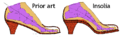 5,782,015 insolia.png 696 × 216; 7 KB
-

-
 Adult and foetal heel bone (calcaneus).jpg 1,942 × 3,281; 400 KB
-
 Arch - Foot (PSF).png 2,003 × 1,256; 58 KB
-

-
 Ballsofthefeet.jpg 820 × 926; 147 KB
-
 Blausen 0411 FootAnatomy eu.png 750 × 1,500; 636 KB
-
 Bones of the feet; two figures. Lithograph by Battistelli af Wellcome V0008194EL.jpg 1,149 × 1,548; 1,019 KB
-
 Bones of the feet; two figures. Lithograph by Battistelli af Wellcome V0008194ER.jpg 1,180 × 1,671; 1.07 MB
-
 Bones of the foot. Colour wood engraving with letterpress Wellcome V0008694.jpg 2,124 × 3,402; 3.5 MB
-
 Braus 1921 303.png 1,090 × 638; 1.99 MB
-
 Braus 1921 311.1.png 1,553 × 1,518; 6.76 MB
-
 Braus 1921 311.2.png 1,469 × 1,520; 6.4 MB
-
 Braus 1921 312.png 1,520 × 1,698; 7.4 MB
-
 Bänder am Fuss - 4686.jpg 2,081 × 4,720; 4.82 MB
-
 Bänder am Fuss - 4716.jpg 1,096 × 2,252; 1.07 MB
-
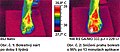
 Cévní obraz - srovnání před a po.jpg 763 × 726; 91 KB
-
-
 Dešinės pėdos jungtys.png 897 × 650; 475 KB
-
 Die Gartenlaube (1855) b 530.jpg 349 × 466; 33 KB
-
 Fasciaplantar-frontal.jpg 1,439 × 2,259; 1.3 MB
-
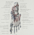
 First metatarsal bone has moved aside1.jpg 444 × 683; 50 KB
-
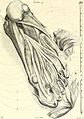
 Foot "Anatomia Humani Corporis", Bidloo, 1685 Wellcome L0014232.jpg 1,186 × 1,690; 759 KB
-
 Foot Arche (PSF).png 2,053 × 1,357; 348 KB
-

-
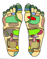
 Foot Chart1 small.png 249 × 314; 26 KB
-
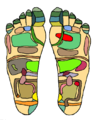
 Foot chart1.png 554 × 697; 205 KB
-
 Foot of a 13 year old - 4688.jpg 747 × 1,818; 901 KB
-
 Foot retro.JPG 2,288 × 1,712; 1.35 MB
-
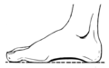
 Foot.png 214 × 356; 10 KB
-
 Gray1239 Arabic YM.jpg 3,496 × 3,912; 2.02 MB
-
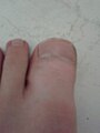 Hallux Romanux 2.jpg 1,920 × 2,560; 1.21 MB
-
 Hallux Romanux.jpg 1,920 × 2,560; 1.15 MB
-
 Homo footprints.jpg 1,552 × 928; 153 KB
-
 Huesos hallux.jpg 766 × 336; 39 KB
-
 Huesos hallux2.jpg 225 × 801; 55 KB
-
 MNH - Mumie Fuß.jpg 3,390 × 1,998; 1.41 MB
-
 Muscles- feet and hand Wellcome L0006624.jpg 1,136 × 1,751; 495 KB
-
 Nerfs de la face plantaire du pied droit.png 591 × 742; 166 KB
-
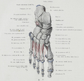
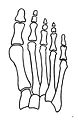 Normal foot skeleton1.jpg 452 × 693; 52 KB
-
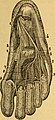
 NormaleFusssohle mit eingezeichbetemSkelett undBodenflache.gif 1,119 × 2,919; 127 KB
-
 Obr12.jpg 380 × 166; 117 KB
-
-
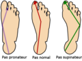 Pas pronateur-supinateur-normal.png 400 × 297; 40 KB
-
 PF-PainAreas-ar.jpg 742 × 875; 202 KB
-
 PF-PainAreas.jpg 742 × 875; 62 KB
-
 PF-PlantarMove.jpg 622 × 866; 55 KB
-
 Plantar Bewegung.gif 524 × 720; 54 KB
-
 Plantar Cutaneous Nerves.png 162 × 104; 36 KB
-
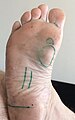 Plantar fibromatosis.jpg 1,099 × 1,758; 274 KB
-
 Posicion referencia.JPG 768 × 576; 55 KB
-
 PSM V17 D758 Normal and boot deformed feet.jpg 3,806 × 2,869; 701 KB
-
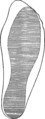
 PSM V24 D664 Well formed healthy foot.jpg 1,008 × 1,585; 224 KB
-
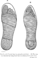
 PSM V24 D666 Outlines of a normal foot and a cramping shoe.jpg 1,678 × 623; 105 KB
-
 Röntgen, Hammerzeh am mittleren Zeh.jpg 769 × 1,259; 203 KB
-
 Science ofDressTo face p239.png 1,493 × 3,072; 501 KB
-
 Science ofDressTo face p239cut.png 1,157 × 3,072; 360 KB
-
 Science ofDressTo face p240.png 2,000 × 3,072; 1.11 MB
-
 Science ofDressTo face p240cut.png 2,200 × 3,072; 1.02 MB
-
 Science ofDressTo face p240cutA.png 1,001 × 3,072; 769 KB
-
 Science ofDressTo face p240cutB.png 1,058 × 3,072; 738 KB
-
 Sehne Musculus extensor hallucis longus - Sono laengs zusammengesetzt - 001.png 1,155 × 291; 185 KB
-
 Slide2ALE.JPG 960 × 720; 80 KB
-
 Soldiersfootmili00munsrich Fig7 anteroposterior arch.png 1,012 × 572; 61 KB
-
 Text-book of operative surgery (1903) (14762066304).jpg 2,329 × 3,697; 1.4 MB
-
 The bones of the foot, from Camper, Dissertation ... Wellcome L0004215.jpg 1,280 × 1,452; 966 KB
-
 TPedBar6.jpg 200 × 396; 9 KB
-
 UBC-BME-KNT-20-008.png 756 × 588; 114 KB
-
 Wax(?) model of a dissected foot, showing tendons. Pencil an Wellcome V0008281.jpg 2,700 × 2,651; 3.69 MB
-

_(14597413648).jpg)
.jpg)
.png)
_(14754215121).jpg)


_(14596371460).jpg)

_b_530.jpg)


.png)


_(14801787723).jpg)


_(14762066304).jpg)


_model_of_a_dissected_foot,_showing_tendons._Pencil_an_Wellcome_V0008281.jpg)
_v_Praze_(ilustracni_fotografie)_(UK0242).jpg)