File:Wound healing phases.svg
Jump to navigation
Jump to search

Size of this PNG preview of this SVG file: 713 × 139 pixels. Other resolutions: 320 × 62 pixels | 640 × 125 pixels | 1,024 × 200 pixels | 1,280 × 250 pixels | 2,560 × 499 pixels.
Original file (SVG file, nominally 713 × 139 pixels, file size: 439 KB)
Captions
Captions
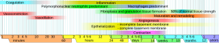
Phases of wound healing and processing
Summary[edit]
| Description |
English: Phases of wound healing. Limits vary within faded intervals, mainly by wound severity. |
| Date | |
| Source | Own work (from the template Logarithmic time scale - milliseconds to years.svg) |
| Author | Mikael Häggström |
| Other versions |
 |
References[edit]
Inflammation and maturation and remodeling:
- worldwidewounds.com > Figure 3 - The time relationship between the different processes of wound healing. by Gregory S Schultz, Glenn Ladwig and Annette Wysocki - in turn adapted from Asmussen PD, Sollner B. Mechanism of wound healing. In: Wound Care. Tutorial Medical Series. Stuttgart: Hippokrates Verlag, 1993.
Vasoconstriction and vasodilation:
- Stadelmann W.K., Digenis A.G. and Tobin G.R. (1998). Physiology and healing dynamics of chronic cutaneous wounds. The American Journal of Surgery 176 (2): 26S-38S. PMID 9777970 Hamilton, Ont. B.C. Decker, Inc. Electronic book
Angiogenesis:
- Nguyen, D.T., Orgill D.P., Murphy G.F. (2009). Chapter 4: The Pathophysiologic Basis for Wound Healing and Cutaneous Regeneration. Biomaterials For Treating Skin Loss. CRC Press (US) & Woodhead Publishing (UK/Europe), Boca Raton/Cambridge, p. 25-57. (ISBN 978-1-4200-9989-9 Invalid ISBN, ISBN 978-1-84569-363-3)
Polymorphonuclear neutrophils and fibroplasia and granulation tissue formation:
- de la Torre J., Sholar A. (2006). Wound healing: Chronic wounds. Emedicine.com. Accessed January 20, 2008. http://www.emedicine.com/plastic/topic477.htm
Macrophages:
- Expert Reviews in Molecular Medicine. (2003). The phases of cutaneous wound healing. 5: 1. Cambridge University Press. Accessed January 20, 2008. http://www-ermm.cbcu.cam.ac.uk/03005829a.pdf
Fibroblast proliferation and migration
- Stadelmann W.K., Digenis A.G. and Tobin G.R. (1998). Physiology and healing dynamics of chronic cutaneous wounds. The American Journal of Surgery 176 (2): 26S-38S. PMID 9777970 Hamilton, Ont. B.C. Decker, Inc. Electronic book
Collagen deposition, epithelialization and contraction:
- Romo T. and Pearson J.M. 2005. Wound Healing, Skin. Emedicine.com. Accessed December 27, 2006.
Additional note on contraction:
- Mulvaney M. and Harrington A. 1994. Chapter 7: Cutaneous trauma and its treatment. In, Textbook of Military Medicine: Military Dermatology. Office of the Surgeon General, Department of the Army. Virtual Naval Hospital Project. Accessed through web archive on September 15, 2007. https://web.archive.org/web/20031218072356/http://www.vnh.org/MilitaryDerm/Ch7.pdf
Percentage of normal tissue strength:
- Mercandetti M., Cohen A.J. (2005). Wound Healing: Healing and Repair. Emedicine.com. Accessed January 20, 2008. http://www.emedicine.com/plastic/topic411.htm
Upper limit of angiogenesis:
- "Some months" given in: Page 550 (section Angiogenesis, Macrophages, and Remodeling) in: William D. Figg (2008) Angiogenesis an integrative approach from science to medicine, Berlin: Springer ISBN: 0-387-71517-7. [1]
Licensing[edit]
| I, the copyright holder of this work, release this work into the public domain. This applies worldwide. In some countries this may not be legally possible; if so: I grant anyone the right to use this work for any purpose, without any conditions, unless such conditions are required by law. |
File history
Click on a date/time to view the file as it appeared at that time.
| Date/Time | Thumbnail | Dimensions | User | Comment | |
|---|---|---|---|---|---|
| current | 05:22, 18 January 2011 | 713 × 139 (439 KB) | Mikael Häggström (talk | contribs) | Moved info in infobox to image page instead. | |
| 04:52, 14 November 2010 | 713 × 139 (442 KB) | Mikael Häggström (talk | contribs) | simplified legend | ||
| 04:49, 14 November 2010 | 713 × 139 (443 KB) | Mikael Häggström (talk | contribs) | another update | ||
| 15:05, 13 November 2010 | 713 × 139 (443 KB) | Mikael Häggström (talk | contribs) | Removed details about constituents. See sections on granulation tissue formation and remodeling for such details. | ||
| 13:53, 13 November 2010 | 713 × 147 (445 KB) | Mikael Häggström (talk | contribs) | minor adjustment | ||
| 13:47, 13 November 2010 | 713 × 147 (445 KB) | Mikael Häggström (talk | contribs) | Distinguished collagen types | ||
| 18:53, 3 November 2010 | 725 × 136 (443 KB) | Mikael Häggström (talk | contribs) | Removed redundant infobox | ||
| 05:01, 21 October 2010 | 746 × 136 (445 KB) | Mikael Häggström (talk | contribs) | Delimited angiogenesis | ||
| 04:44, 21 October 2010 | 746 × 136 (445 KB) | Mikael Häggström (talk | contribs) | minor scale adjustment | ||
| 14:30, 7 October 2010 | 746 × 136 (453 KB) | Mikael Häggström (talk | contribs) | moved infobox |
You cannot overwrite this file.
File usage on Commons
The following 3 pages use this file: