File:Uterine anatomy..jpg
Jump to navigation
Jump to search
No higher resolution available.
Uterine_anatomy..jpg (800 × 320 pixels, file size: 82 KB, MIME type: image/jpeg)
Captions
Captions
Add a one-line explanation of what this file represents
| Description |
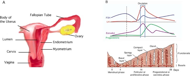
English: The female reproductive tract is shown in panel A comprising a cervix, uterus, and Fallopian tubes. Histologically, the uterine wall can be subdivided into the outer perimetrium, inner endometrium, and intermediate myometrium. Panel B shows cyclic changes during the menstrual cycle. In the sexually mature woman, the uterus goes through monthly cycles in which the functionalis portion of the endometrial lining of the uterus is stimulated to grow by estrogen, which is produced by the ovary. After ovulation, progesterone is also produced by the ovary causing the lining of the uterus to stop growing. If pregnancy is not established, the functionalis is mostly shed, i.e. menstruation. |
| Date | Published September 30, 2008. |
| Source |
[1] Direct
|
| Author | Teixeira, J., Rueda, B.R., and Pru, J.K., Uterine Stem cells (September 30, 2008), StemBook, ed. The Stem Cell Research Community, StemBook, doi/10.3824/stembook.1.16.1, http://www.stembook.org. |
| Permission (Reusing this file) |
This file is licensed under the Creative Commons Attribution 3.0 Unported license.
|
File history
Click on a date/time to view the file as it appeared at that time.
| Date/Time | Thumbnail | Dimensions | User | Comment | |
|---|---|---|---|---|---|
| current | 18:56, 5 April 2013 | 800 × 320 (82 KB) | Smallbot (talk | contribs) | Uploading CC-BY images from the the StemBook http://www.stembook.org/ 108/173 |
You cannot overwrite this file.
File usage on Commons
There are no pages that use this file.
File usage on other wikis
The following other wikis use this file:
- Usage on en.wikipedia.org
- Usage on sc.wikipedia.org