File:Transfer cells fpls-04-00221-g001.jpg
Jump to navigation
Jump to search

No higher resolution available.
Transfer_cells_fpls-04-00221-g001.jpg (394 × 428 pixels, file size: 174 KB, MIME type: image/jpeg)
Captions
Captions
Add a one-line explanation of what this file represents
Summary[edit]
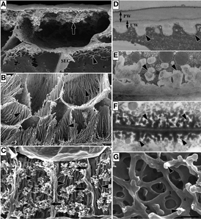
| Description | Figure 1. Images of transfer cells of developing seeds during their storage phase illustrating ingrowth wall morphologies. (A–C) Scanning (A,B) and field emission scanning (C) electron microscope images of cells following freeze-fracture, removal of their cytoplasm and fixation [for method see Talbot et al. (2002)]. (A) Epidermal transfer cells (ETC) of a Vicia faba cotyledon with an extensive reticulate ingrowth wall labyrinth (arrow) polarized to the outer periclinal wall. Ingrowth wall deposition (dart) is restricted to wall portions abutting intercellular spaces adjacent to the sub-epidermal cells (SEC) [modified after Talbot et al. (2001)]. (B) Basal endosperm transfer cells of Zea mays exhibiting flange wall ingrowth morphology [modified after Talbot et al. (2002)]. The wall ingrowth ribs (darts) extend the length of each cell and are more extensive at their outer periclinal walls. (C) Thin-walled parenchyma transfer cells located at the inner surface of the inner seed coat of Gossypium hirsutum with wall ingrowth flanges (darts) extending the length of each cell on which are deposited groups of reticulate wall ingrowths (arrows) [modified after Pugh et al. (2010)]. (D–F) Transmission electron microscope images of portions of transverse sections of transfer cells: (D) The outer periclinal wall of an adaxial epidermal cell of a V. faba cotyledon induced to trans-differentiate to a transfer cell morphology. A uniform wall (UW), distinguishable from the original primary wall (PW) by a different electron opaqueness, is deposited against the primary wall and small papillate wall ingrowths (darts) arise from it. (E) Small papillate ingrowths (darts) of a seed coat transfer cell of V. faba exhibiting reticulate architecture. (F) Antler-shaped reticulate wall ingrowths (darts) of a nucellar projection transfer cell of a developing Triticum turgidum var. durum seed [modified after Wang et al. (1994)]. (G) Field emission scanning electron microscope image of the cytoplasmic face of the reticulate ingrowth wall labyrinth of an abaxial epidermal transfer cell of a V. faba cotyledon following removal of the cytoplasm and dry cleaving [for method see Talbot et al. (2001), image modified after Talbot et al. (2001)]. Note the multi-layered fenestrated sheets of wall material (numbered) and the small wall ingrowth papillae arising from the most recently deposited layer (darts). Single scale bar for (A,B) = 2.5 μm; for (C) = 5 μm; for (D,E) = 1 μm; for (F) = 0.25 μm; for (G) = 0.5 μm. |
| Date | |
| Source | https://www.frontiersin.org/files/Articles/51224/fpls-04-00221-HTML/image_m/fpls-04-00221-g001.jpg Intersection of transfer cells with phloem biology—broad evolutionary trends, function, and induction, Front. Plant Sci., 01 July 2013, Sec. Plant Physiology, Volume 4 - 2013, https://doi.org/10.3389/fpls.2013.00221 |
| Author | Felicity A. Andriunas, Hui-Ming Zhang, Xue Xia, John W. Patrick, Christina E. Offler |
Open access
Licensing[edit]
This file is licensed under the Creative Commons Attribution 3.0 Unported license.
- You are free:
- to share – to copy, distribute and transmit the work
- to remix – to adapt the work
- Under the following conditions:
- attribution – You must give appropriate credit, provide a link to the license, and indicate if changes were made. You may do so in any reasonable manner, but not in any way that suggests the licensor endorses you or your use.
File history
Click on a date/time to view the file as it appeared at that time.
| Date/Time | Thumbnail | Dimensions | User | Comment | |
|---|---|---|---|---|---|
| current | 03:25, 1 January 2024 | | 394 × 428 (174 KB) | Rasbak (talk | contribs) | {Information |description=Figure 1. Images of transfer cells of developing seeds during their storage phase illustrating ingrowth wall morphologies. (A–C) Scanning (A,B) and field emission scanning (C) electron microscope images of cells following freeze-fracture, removal of their cytoplasm and fixation [for method see Talbot et al. (2002)]. (A) Epidermal transfer cells (ETC) of a Vicia faba cotyledon with an extensive reticulate ingrowth wall labyrinth (arrow) polarized to the outer periclin... |
You cannot overwrite this file.
File usage on Commons
There are no pages that use this file.
File usage on other wikis
The following other wikis use this file:
- Usage on nl.wikipedia.org