File:Thygeson's superficial punctate keratitis.jpg
Jump to navigation
Jump to search

Size of this preview: 800 × 533 pixels. Other resolutions: 320 × 213 pixels | 640 × 427 pixels | 1,024 × 683 pixels | 1,200 × 800 pixels.
Original file (1,200 × 800 pixels, file size: 127 KB, MIME type: image/jpeg)
Captions
Captions
Add a one-line explanation of what this file represents
| Description |
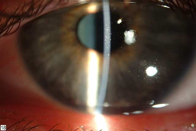
English: Left cornea before cyclosporin A treatment. Note the slightly elevated, grayish-white subepithelial opacities. Clinical appearance was similar in the right eye.Hasanreisoglu and Avisar Cases Journal 2008 1:415 doi:10.1186/1757-1626-1-415 Русский: Поверхностный точечный кератит Тайджесона (иногда "Тигесона": Thygeson). Чуть выдающиеся серовато-белые субэпителиальные помутнения. Смотри также: та же роговица после лечения циклоспорином. |
| Date | |
| Source | Long-term topical cyclosporin A therapy in Thygeson's superficial punctate keratitis: a case report |
| Author | Murat Hasanreisoglu and Rahamim Avisar |
| Permission (Reusing this file) |
© 2008 Hasanreisoglu and Avisar; licensee BioMed Central Ltd. This is an Open Access article distributed under the terms of the Creative Commons Attribution License (https://creativecommons.org/licenses/by/2.0), which permits unrestricted use, distribution, and reproduction in any medium, provided the original work is properly cited. |
This file is licensed under the Creative Commons Attribution 2.0 Generic license.
- You are free:
- to share – to copy, distribute and transmit the work
- to remix – to adapt the work
- Under the following conditions:
- attribution – You must give appropriate credit, provide a link to the license, and indicate if changes were made. You may do so in any reasonable manner, but not in any way that suggests the licensor endorses you or your use.
| Annotations | This image is annotated: View the annotations at Commons |
File history
Click on a date/time to view the file as it appeared at that time.
| Date/Time | Thumbnail | Dimensions | User | Comment | |
|---|---|---|---|---|---|
| current | 13:18, 13 August 2009 | | 1,200 × 800 (127 KB) | CopperKettle (talk | contribs) | {{Information |Description={{en|1=Left cornea before cyclosporin A treatment. Note the slightly elevated, grayish-white subepithelial opacities. Clinical appearance was similar in the right eye.Hasanreisoglu and Avisar Cases Journal 2008 1:415 doi:10.11 |
You cannot overwrite this file.
File usage on Commons
The following 2 pages use this file:
File usage on other wikis
The following other wikis use this file:
- Usage on ar.wikipedia.org
- Usage on de.wikipedia.org
- Usage on es.wikipedia.org
- Usage on hr.wikipedia.org
- Usage on it.wikipedia.org
- Usage on ru.wikipedia.org
- Usage on sh.wikipedia.org
- Usage on tt.wikipedia.org
- Usage on www.wikidata.org