File:Thin section scan crossed polarizers Siilinjärvi R636-116.75.jpg

Original file (4,010 × 2,100 pixels, file size: 2.21 MB, MIME type: image/jpeg)
Captions
Captions
Summary[edit]
| Description |
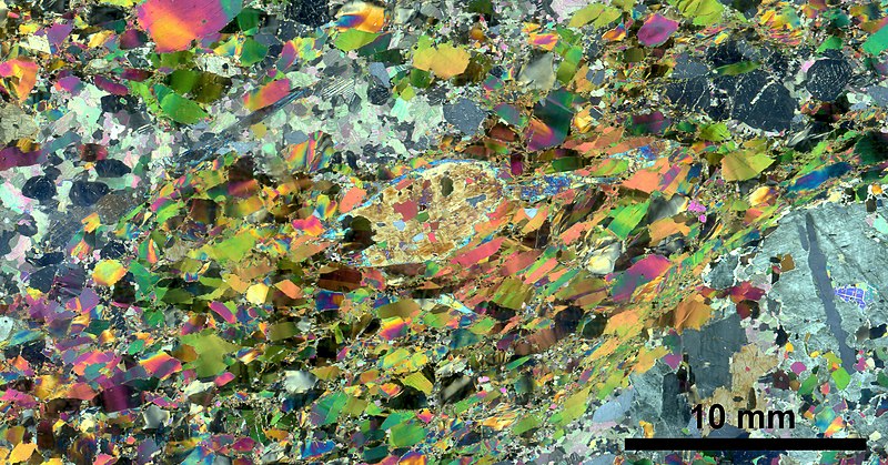
 English: Scanned image of thin section from Siilinjärvi apatite ore in cross polarised transmitted light.
The thin section has a very low amount of small mica chips. The bigger mica flakes in the sample are oriented, but shearing has not affected the grain size nor formed any foliation. Magnetite is found as accessory mineral in the thin section. Tetraferriphlogopite forms 75 area-% of the sample and is mostly present as somewhat elongated flakes. The grains are usually 1-2 mm long and 0.3-1.5 mm wide. Smaller chips are in minority and do not show any orientation. Amphiboles look often quite broken, but usually the grains still appear large. The blue colour is quite weak, but still prominent. The amphiboles are a mixture between richterite and tremolite. The biggest grains are > 7 mm in diameter and have inclusions of micas and apatites. Some smaller (diameter 0.3-1.5 mm) euhedral grains are also present. Amphiboles form 10 area-% of the thin section. Carbonates look quite pure in the thin section and they are grown into large mineral aggregates. The aggregates have other mineral grains in them, mostly apatite. Carbonate grain size varies a lot, and the biggest grains are usually found in the middle of the granular aggregates (diameter almost 2 mm). The aggregate margins feature smaller grains. Carbonates are also quite common as small individual grains in the mica groundmass. Calcite is more common carbonate mineral in the aggregates, dolomite as individual grains. Carbonates form 8 area-% of the thin section. Apatite is found in companion with carbonates and forms 7 area-% of the thin section. Mostly the apatite is present as big rounded grains together with carbonates, but there is also a lot of small apatite pieces in the mica groundmass. Average sized apatite is 1-3 mm in diameter, the pieces in the groundmass are 0.1-1 mm in diameter. |
| Date | |
| Source | Own work |
| Author | kallerna |
| Other versions |
|


This image has been assessed using the Quality image guidelines and is considered a Quality image.
|
| This image was uploaded as part of Wiki Science Competition 2019. |
Licensing[edit]
- You are free:
- to share – to copy, distribute and transmit the work
- to remix – to adapt the work
- Under the following conditions:
- attribution – You must give appropriate credit, provide a link to the license, and indicate if changes were made. You may do so in any reasonable manner, but not in any way that suggests the licensor endorses you or your use.
- share alike – If you remix, transform, or build upon the material, you must distribute your contributions under the same or compatible license as the original.
File history
Click on a date/time to view the file as it appeared at that time.
| Date/Time | Thumbnail | Dimensions | User | Comment | |
|---|---|---|---|---|---|
| current | 19:05, 12 November 2019 | | 4,010 × 2,100 (2.21 MB) | Kallerna (talk | contribs) | User created page with UploadWizard |
You cannot overwrite this file.
File usage on Commons
The following 27 pages use this file:
- Image sets from Wiki Science Competition 2019 in the rest of the world
- User talk:Kallerna/2015-2019
- Commons:Quality images/Subject/Microscopic
- Commons:Quality images candidates/Archives November 17 2019
- Commons:Wiki Science Competition 2019/Winners/Image sets
- Commons:Wiki Science Competition 2019/Winners/Image sets/round 2
- Commons:Wiki Science Competition 2019/Winners/International
- File:Thin section microscopy Siilinjärvi R636 116 etched.jpg
- File:Thin section scan crossed polarizers Siilinjärvi 501-M3.jpg
- File:Thin section scan crossed polarizers Siilinjärvi H10.jpg
- File:Thin section scan crossed polarizers Siilinjärvi H16.jpg
- File:Thin section scan crossed polarizers Siilinjärvi H2.jpg
- File:Thin section scan crossed polarizers Siilinjärvi R276-108.40.jpg
- File:Thin section scan crossed polarizers Siilinjärvi R301-61.70.jpg
- File:Thin section scan crossed polarizers Siilinjärvi R616-78.20.jpg
- File:Thin section scan crossed polarizers Siilinjärvi R625-110.80.jpg
- File:Thin section scan crossed polarizers Siilinjärvi R625-122.05.jpg
- File:Thin section scan crossed polarizers Siilinjärvi R625-145.40.jpg
- File:Thin section scan crossed polarizers Siilinjärvi R625-153.25.jpg
- File:Thin section scan crossed polarizers Siilinjärvi R625-47.40.jpg
- File:Thin section scan crossed polarizers Siilinjärvi R636-105.90.jpg
- File:Thin section scan crossed polarizers Siilinjärvi R636-116.75.jpg
- File:Thin section scan crossed polarizers Siilinjärvi R636-120.45.jpg
- File:Thin section scan crossed polarizers Siilinjärvi R636-131.20.jpg
- File:Thin section scan crossed polarizers Siilinjärvi R636-136.45.jpg
- File:Thin section scan crossed polarizers Siilinjärvi R636-42.60.jpg
- File:Thin section scan crossed polarizers Siilinjärvi R636-63.35.jpg
File usage on other wikis
The following other wikis use this file:
- Usage on ca.wikipedia.org
- Usage on en.wikipedia.org
- Usage on es.wikipedia.org
- Usage on fr.wiktionary.org
- Usage on ru.wikipedia.org
- Usage on uk.wikipedia.org