File:Red on centre green off centre.png
Jump to navigation
Jump to search
No higher resolution available.
Red_on_centre_green_off_centre.png (634 × 474 pixels, file size: 48 KB, MIME type: image/png)
Captions
Captions
Add a one-line explanation of what this file represents
Summary[edit]
| Description |
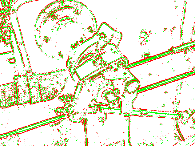
English: An example of edge detection using an emulation of retinal receptive fields. This is what the output of the optic nerve would look like (as a very rough approximation: the fovea is an almost 1:1 high definition visual area and the receptive fields increase in size from the centre of the retina to the peripheral). This image was processed in Java using a 52-pixel template (contact me for source code). Both types (on centre and off centre) cells are shown in different colours. Here is the original preprocessed image.
Русский: Распознавание границ изображения (краёв, углов) рецептивными полями сетчатки (грубая компьютерная аппроксимация). Размеры рецептивных полей увеличиваются от центра сетчатки к её периферии). Визуальная информация от двух типов клеток (с on- и off-центрами) показана красным и зелёным цветом, соответственно.
Автор изображения – User:Simpsons_contributor |
| Date | |
| Source | originally uploaded to the English language wikipedia |
| Author | Own work by Simpsons contributor |
Licensing[edit]
| This work has been released into the public domain by its author, Simpsons contributor at English Wikipedia. This applies worldwide. In some countries this may not be legally possible; if so: Simpsons contributor grants anyone the right to use this work for any purpose, without any conditions, unless such conditions are required by law. |
Original upload log[edit]
The original description page was on en.wikipedia (file log). All following user names refer to en.wikipedia.
- 08:52, 31 May 2009 (UTC) Simpsons contributor 634×474 (48 KB) ({{Information |Description = An example of edge detection using an emulation of retinal receptive fields. This is what the output of the optic nerve would look like (as a very rough approximation: the fovea is an almost 1:1 high definition visual area)
File history
Click on a date/time to view the file as it appeared at that time.
| Date/Time | Thumbnail | Dimensions | User | Comment | |
|---|---|---|---|---|---|
| current | 08:51, 21 March 2011 | | 634 × 474 (48 KB) | JeanneMish (talk | contribs) | {{Information |Description ={{en|1=An example of edge detection using an emulation of retinal receptive fields. This is what the output of the optic nerve would look like (as a very rough approximation: the fovea is an almost 1:1 high definition visual |
You cannot overwrite this file.
File usage on Commons
The following page uses this file:
- File:Red on centre green off centre .png (file redirect)
File usage on other wikis
The following other wikis use this file:
- Usage on en.wikipedia.org
- Usage on fr.wikipedia.org
- Usage on ru.wikipedia.org