File:Ptetraurelia C-basales.jpg
Ptetraurelia_C-basales.jpg (358 × 212 pixels, file size: 46 KB, MIME type: image/jpeg)
Captions
Captions
Summary[edit]
| Description |
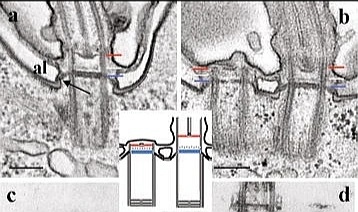
Español: Fig. 3. Secciones longitudinales a través de los cuerpos basales de Paramecium. a) conexión entre el Tz y la superficie celular. Dentro del cuerpo basal, el Tz está organizado en tres placas: la placa terminal (línea azul), la placa intermedia (blanca) y la placa axosómica (línea roja). |
| Date | |
| Source | Paramecium tetraurelia basal body structure. Cilia vol 5 núm 6 |
| Author | Tassin A.M., Lemullois M., Aubusson-Fleury A. |
| Permission (Reusing this file) |
From the article's Rights and Permissions section: Open Access This article is distributed under the terms of the Creative Commons Attribution 4.0 International License (https://creativecommons.org/licenses/by/4.0/), which permits unrestricted use, distribution, and reproduction in any medium, provided you give appropriate credit to the original author(s) and the source, provide a link to the Creative Commons license, and indicate if changes were made. The Creative Commons Public Domain Dedication waiver (https://creativecommons.org/publicdomain/zero/1.0/) applies to the data made available in this article, unless otherwise stated. |
Licensing[edit]
- You are free:
- to share – to copy, distribute and transmit the work
- to remix – to adapt the work
- Under the following conditions:
- attribution – You must give appropriate credit, provide a link to the license, and indicate if changes were made. You may do so in any reasonable manner, but not in any way that suggests the licensor endorses you or your use.
File history
Click on a date/time to view the file as it appeared at that time.
| Date/Time | Thumbnail | Dimensions | User | Comment | |
|---|---|---|---|---|---|
| current | 03:59, 21 April 2020 | | 358 × 212 (46 KB) | Sanador2.0 (talk | contribs) | {{Information |description ={{es|1=Fig. 3. Secciones longitudinales a través de los cuerpos basales de Paramecium.<br> a) conexión entre el Tz y la superficie celular. Dentro del cuerpo basal, el Tz está organizado en tres placas: la placa terminal (línea azul), la placa intermedia (blanca) y la placa axosómica (línea roja).<br> Fuera del cuerpo basal, la placa terminal se extiende para unir el epiplasma (flecha).<br> Dentro del cuerpo basal, se observan gránulos densos. (Al) saco alveolar... |
You cannot overwrite this file.
File usage on Commons
There are no pages that use this file.
File usage on other wikis
The following other wikis use this file:
- Usage on es.wikipedia.org