File:Proceedings of the Washington Academy of Sciences. Volume 3. Plate IX.png
Jump to navigation
Jump to search

Size of this preview: 371 × 599 pixels. Other resolutions: 148 × 240 pixels | 297 × 480 pixels | 476 × 768 pixels | 634 × 1,024 pixels | 2,099 × 3,388 pixels.
Original file (2,099 × 3,388 pixels, file size: 4.03 MB, MIME type: image/png)
Captions
Captions
Add a one-line explanation of what this file represents
Summary[edit]
| Description |
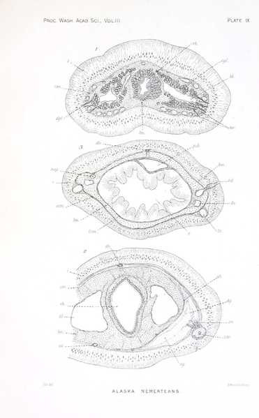
English: Carinella speciosa (=Tubulanus polymorphus; Nemertea: Palaeonemertea: Tubulanidae).
|
| Date | |
| Source | Coe, W. R. (1901). Papers from the Harriman Alaska Expedition. XX. Nemerteans. 3: 1–110. Plate IX in the Biodiversity Heritage Library |
| Author | Wesley Roswell Coe (1869–1960) |
| Other versions |
.png)
.png)
.png)
Licensing[edit]
This media file is in the public domain in the United States. This applies to U.S. works where the copyright has expired, often because its first publication occurred prior to January 1, 1929, and if not then due to lack of notice or renewal. See this page for further explanation.
|
 | |
 |
This image might not be in the public domain outside of the United States; this especially applies in the countries and areas that do not apply the rule of the shorter term for US works, such as Canada, Mainland China (not Hong Kong or Macao), Germany, Mexico, and Switzerland. The creator and year of publication are essential information and must be provided. See Wikipedia:Public domain and Wikipedia:Copyrights for more details.
|
File history
Click on a date/time to view the file as it appeared at that time.
| Date/Time | Thumbnail | Dimensions | User | Comment | |
|---|---|---|---|---|---|
| current | 00:19, 22 July 2014 | | 2,099 × 3,388 (4.03 MB) | Mithril (talk | contribs) | =={{int:filedesc}}== {{Information |Description = {{en|1=Ribbon worms from Proceedings of the Washington Academy of Sciences, volume 3.}} |Source = Coe, W. R. (1901). Papers from the Harriman Alaska Expedition. XX. Nemerteans. '''3''': 1–110. [h... |
You cannot overwrite this file.
File usage on Commons
The following 3 pages use this file: