File:Phragmosome.svg
Jump to navigation
Jump to search

Size of this PNG preview of this SVG file: 369 × 467 pixels. Other resolutions: 190 × 240 pixels | 379 × 480 pixels | 607 × 768 pixels | 809 × 1,024 pixels | 1,618 × 2,048 pixels.
Original file (SVG file, nominally 369 × 467 pixels, file size: 33 KB)
Captions
Captions
Add a one-line explanation of what this file represents
Summary
[edit]
| Description |
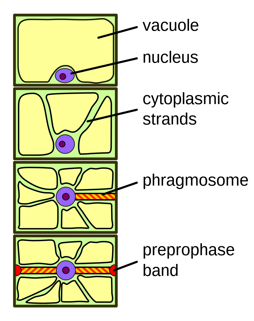
English: Phragmosome formation in a highly vacuolated plant cell. From top to bottom: 1) Interphase cell with large central vacuole. 2) Cytoplasmic strands starting to penetrate vacuole. 3) Nucleus migration into center and formation of the phragmosome. 4) Phragmosome formation completed and formation of preprophase band marking future cell division plane. When mitosis is completed, the new cell wall will form starting from the center along the plane occupied by the phragmosome. |
| Date | (UTC) |
| Source | http://en.wikipedia.org/wiki/Image:Phragmosome.png |
| Author | en:User:Tameeria (original); Pbroks13 (talk) (redraw) |
Licensing
[edit]
This file is licensed under the Creative Commons Attribution 3.0 Unported license.
- You are free:
- to share – to copy, distribute and transmit the work
- to remix – to adapt the work
- Under the following conditions:
- attribution – You must give appropriate credit, provide a link to the license, and indicate if changes were made. You may do so in any reasonable manner, but not in any way that suggests the licensor endorses you or your use.
File history
Click on a date/time to view the file as it appeared at that time.
| Date/Time | Thumbnail | Dimensions | User | Comment | |
|---|---|---|---|---|---|
| current | 05:53, 13 August 2008 | | 369 × 467 (33 KB) | Pbrks (talk | contribs) | {{Information |Description={{en|1=Phragmosome formation in a highly vacuolated plant cell. From top to bottom: 1) Interphase cell with large central vacuole. 2) Cytoplasmic strands starting to penetrate vacuole. 3) Nucleus migration into center and format |
You cannot overwrite this file.
File usage on Commons
The following 2 pages use this file:
File usage on other wikis
The following other wikis use this file:
- Usage on bs.wikipedia.org
- Usage on en.wikipedia.org
- Usage on gl.wikipedia.org
- Usage on he.wikipedia.org
- Usage on hr.wikipedia.org
- Usage on hy.wikipedia.org
- Usage on it.wikipedia.org
- Usage on pt.wikipedia.org