File:Periclimenaeus caraibicus and Hymenocera picta (10.7717-peerj.846) Figure 4.png
Jump to navigation
Jump to search

Size of this preview: 523 × 599 pixels. Other resolutions: 209 × 240 pixels | 419 × 480 pixels | 670 × 768 pixels | 894 × 1,024 pixels | 2,246 × 2,573 pixels.
Original file (2,246 × 2,573 pixels, file size: 6.35 MB, MIME type: image/png)
Captions
Captions
Add a one-line explanation of what this file represents
Summary[edit]
_Figure_4.png&action=edit§ion=1)
| Description |
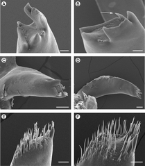
English: Figure 4: Palaemonidae: Periclimenaeus caraibicus and Hymenoceridae: Hymenocera picta.
|
| Date | |
| Source | https://doi.org/10.7717/peerj.846 |
| Author | Ashelby CW, De Grave S, Johnson ML. 2015. Preliminary observations on the mandibles of palaemonoid shrimp (Crustacea: Decapoda: Caridea: Palaemonoidea) PeerJ 3:e846 https://doi.org/10.7717/peerj.846 |
| Permission (Reusing this file) |
This file is licensed under the Creative Commons Attribution 4.0 International license.
|
File history
Click on a date/time to view the file as it appeared at that time.
| Date/Time | Thumbnail | Dimensions | User | Comment | |
|---|---|---|---|---|---|
| current | 12:09, 3 November 2020 | | 2,246 × 2,573 (6.35 MB) | Christian Ferrer (talk | contribs) | {{Information | description = {{en|1= Figure 4: Palaemonidae: ''Periclimenaeus caraibicus'' and Hymenoceridae: ''Hymenocera picta''. :Palaemonidae (Pontoniinae): Periclimenaeus caraibicus, (A) pars molaris of right mandible; (B) pars molaris of right mandible (spine-like tuft of Type II cuticular structures indicated by white arrow); (C) pars molaris of left mandible. Hymenoceridae: Hymenocera picta, (D) right mandible; (E) distal end of pars molaris of right mandible; (F) distal end of pars... |
You cannot overwrite this file.
File usage on Commons
The following page uses this file:
File usage on other wikis
The following other wikis use this file:
- Usage on ceb.wikipedia.org
_Figure_4.png&oldid=699705925)