File:Oxides particles.jpg

Original file (3,216 × 3,264 pixels, file size: 4.42 MB, MIME type: image/jpeg)
Captions
Captions
Summary[edit]
| Description |
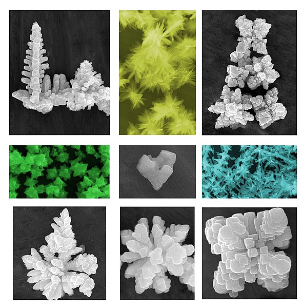
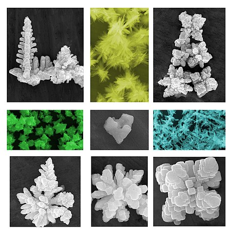
English: Scanning Electron Microscopy (SEM) images showing particles of advanced functional oxides. The powders are synthesized by chemical methods in solution (precipitation, hydrothermal and solvothermal synthesis). Color (if added) helps to emphasize the grayscale levels.
The grayscale images show dendritic growth of barium titanate (BaTiO3) by hydrothermal synthesis. The different morphologies depend on the synthesis conditions: size and shape can be varied by changing the concentration of precursors, the reaction temperature and the time. In general, the synthesis of BaTiO3 by precipitation from aqueous solution allows to produce particles with spherical shape with size that can be tailored, from a few nanometers to several hundred nanometers, by decreasing the concentration of reactants. At very low concentration the particles have the tendency to develop a dendritic-like morphology, as reported in the images.Italiano: Immagini al microscopio elettronico a scansione (SEM) che mostrano particelle di ossidi funzionali avanzati. Le polveri sono sintetizzate con metodi chimici in soluzione (precipitazione, sintesi idrotermale e solvotermale). Il colore (se aggiunto) aiuta a enfatizzare i livelli di di grigio.
Le immagini in scala di grigio mostrano crescita dendritica di titanato di bario (BaTiO3) per sintesi idrotermale. Le diverse morfologie dipendono dalle condizioni di sintesi: dimensioni e forma possono essere modificate cambiando la concentrazione dei precursori, la temperatura e il tempo di reazione. In generale, la sintesi di BaTiO3 mediante precipitazione da soluzione acquosa consente di produrre particelle di forma sferica con dimensioni che possono essere regolate, da pochi nanometri a diverse centinaia di nanometri, diminuendo la concentrazione dei reagenti. In condizioni di concentrazione molto bassa le particelle hanno la tendenza a sviluppare una morfologia dendritica, come riportato nelle immagini. |
| Date | |
| Source | Own work |
| Author | M.T. Buscaglia, ICMATE-CNR |
Licensing[edit]
- You are free:
- to share – to copy, distribute and transmit the work
- to remix – to adapt the work
- Under the following conditions:
- attribution – You must give appropriate credit, provide a link to the license, and indicate if changes were made. You may do so in any reasonable manner, but not in any way that suggests the licensor endorses you or your use.
| This image was uploaded as part of Wiki Science Competition 2019. |
File history
Click on a date/time to view the file as it appeared at that time.
| Date/Time | Thumbnail | Dimensions | User | Comment | |
|---|---|---|---|---|---|
| current | 21:57, 15 December 2019 | | 3,216 × 3,264 (4.42 MB) | Mtbusca (talk | contribs) | User created page with UploadWizard |
You cannot overwrite this file.
File usage on Commons
The following 5 pages use this file:
File usage on other wikis
The following other wikis use this file:
- Usage on bg.wikipedia.org
- Usage on en.wikipedia.org
- Usage on it.wikipedia.org
- Usage on nl.wikipedia.org