File:Notodasus celebensis (10.7717-peerj.7638) Figure 4.png
Jump to navigation
Jump to search
Size of this preview: 462 × 600 pixels. Other resolutions: 185 × 240 pixels | 370 × 480 pixels | 592 × 768 pixels | 789 × 1,024 pixels | 2,438 × 3,164 pixels.
Original file (2,438 × 3,164 pixels, file size: 1.31 MB, MIME type: image/png)
Captions
Captions
Add a one-line explanation of what this file represents
Summary[edit]
_Figure_4.png&action=edit§ion=1)
| Description |
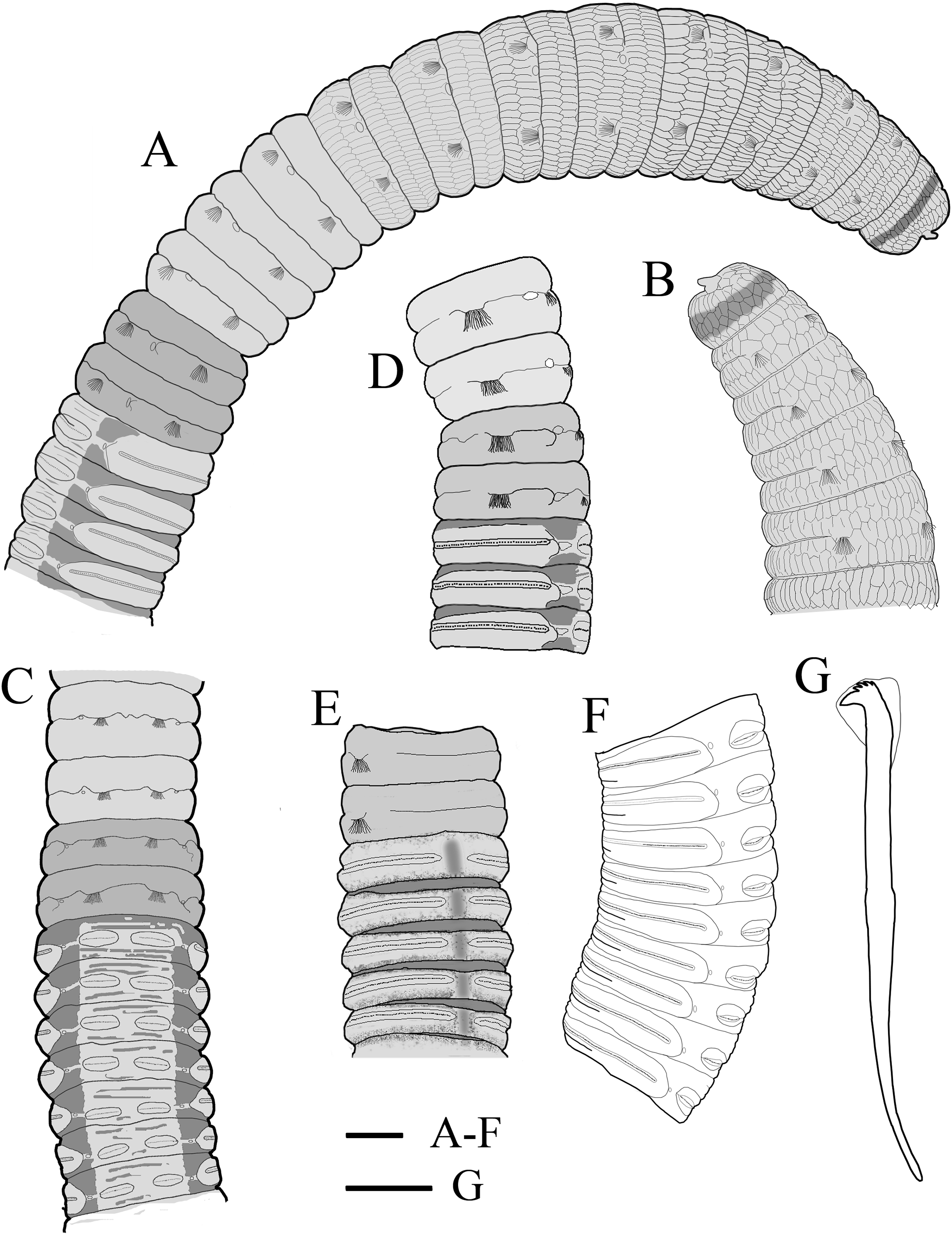
English: Figure 4: Holotype of Notodasus celebensis sp. nov. (TIO-BTS-Poly 101).
|
| Date | |
| Source | https://doi.org/10.7717/peerj.7638 |
| Author | [Drawing by Junhui Lin] Lin J, García-Garza ME, Arbi UY, Wang J. 2019. Two new species of Notodasus Fauchald, 1972 (Annelida: Capitellidae) from the Central Indo-Pacific region. PeerJ 7:e7638 https://doi.org/10.7717/peerj.7638 |
| Permission (Reusing this file) |
This file is licensed under the Creative Commons Attribution 4.0 International license.
|
File history
Click on a date/time to view the file as it appeared at that time.
| Date/Time | Thumbnail | Dimensions | User | Comment | |
|---|---|---|---|---|---|
| current | 19:41, 4 November 2020 | | 2,438 × 3,164 (1.31 MB) | Christian Ferrer (talk | contribs) | {{Information | description = {{en|1= Figure 4: Holotype of ''Notodasus celebensis'' sp. nov. (TIO-BTS-Poly 101). :(A) Anterior 17 chaetigers, lateral view. (B) Anterior end, lateral view. (C) Chaetigers 10–20, dorsal view, showing transition between thorax and abdomen. (D) Chaetigers 10–16, lateral view. (E) Chaetigers 12–18, ventrolateral view. (F) Chaetigers 32–40, lateral view. (G) Neuropodial hook from chaetiger 40. Shading on A–E indicates methyl green staining. Scale bars: A–F, one mm... |
You cannot overwrite this file.
File usage on Commons
There are no pages that use this file.
File usage on other wikis
The following other wikis use this file:
- Usage on ceb.wikipedia.org
- Usage on species.wikimedia.org
_Figure_4.png&oldid=510180523)