File:Mollusca (10.5852-ejt.2021.785.1605) Figure 17.png
Jump to navigation
Jump to search

Size of this preview: 484 × 600 pixels. Other resolutions: 194 × 240 pixels | 387 × 480 pixels | 620 × 768 pixels | 826 × 1,024 pixels | 1,862 × 2,307 pixels.
Original file (1,862 × 2,307 pixels, file size: 3.1 MB, MIME type: image/png)
Captions
Captions
Add a one-line explanation of what this file represents
Summary[edit]
| Description |
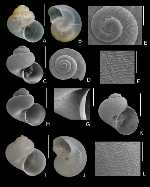
English: Fig. 17. Skeneimorph species 4. A–H. Anekes spiralis Gofas & Luque sp. nov. A–B. Holotype (MNCN 15.05/200139H), BANGAL 0711, V5, 1631 m, diameter 1.7 mm. C–D. SEM micrograph of the holotype. E. Apical view, protoconch and early teleoconch whorl of the holotype. F. Detail of the microsculpture of the adapical part of the last whorl. G. Detail of the umbilical area of the holotype. H. Paratype (MNCN 15.05/200139P), SEM micrograph, same locality, 2.0 mm. I–J. Granigyra inflata (Warén, 1992), BANGAL 0711, V5, 1631 m, diameter 1.8 mm. K–L. Same shell, SEM micrograph and detail of the microsculpture of the last whorl. Scale bars: A–D, H–K = 1 mm; E, G, L = 200 µm; F = 50 µm. |
| Date | |
| Source | https://doi.org/10.5852/ejt.2021.785.1605 |
| Author | Gofas, S., Luque, Ángel A., Oliver, J. D., Templado, J., & Serrano, A. (2021). The Mollusca of Galicia Bank (NE Atlantic Ocean). European Journal of Taxonomy, 785(1), 1–114. |
| Permission (Reusing this file) |
This file is licensed under the Creative Commons Attribution 4.0 International license.
|
| Other versions |
_Figure_17_(cropped).png)
_Figure_17_(cropped).png)
_Figure_17.png&action=edit§ion=1)
File history
Click on a date/time to view the file as it appeared at that time.
| Date/Time | Thumbnail | Dimensions | User | Comment | |
|---|---|---|---|---|---|
| current | 16:43, 27 January 2022 | | 1,862 × 2,307 (3.1 MB) | Christian Ferrer (talk | contribs) | {{Information | description = {{en|1=Fig. 17. Skeneimorph species 4. A–H. ''Anekes spiralis'' Gofas & Luque sp. nov. A–B. Holotype (MNCN 15.05/200139H), BANGAL 0711, V5, 1631 m, diameter 1.7 mm. C–D. SEM micrograph of the holotype. E. Apical view, protoconch and early teleoconch whorl of the holotype. F. Detail of the microsculpture of the adapical part of the last whorl. G. Detail of the umbilical area of the holotype. H. Paratype (MNCN 15.05/200139P), SEM micrograph, same locality, 2.0 mm.... |
You cannot overwrite this file.
File usage on Commons
The following 2 pages use this file:
_Figure_17.png&oldid=625726812)