File:Liver bud morphogenesis..jpg
Jump to navigation
Jump to search
No higher resolution available.
Liver_bud_morphogenesis..jpg (600 × 268 pixels, file size: 93 KB, MIME type: image/jpeg)
Captions
Captions
Add a one-line explanation of what this file represents
| Description |
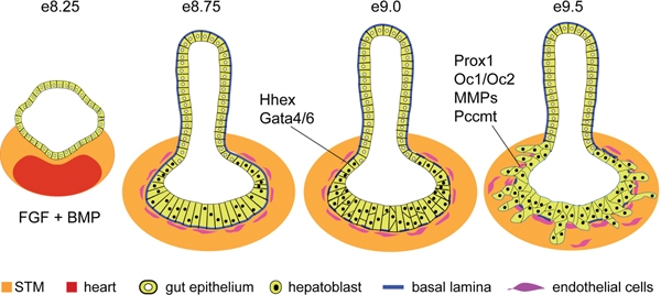
English: The schematics depict transverse sections through the gut tube at the level of the liver diverticulum at different developmental stages in liver bud formation. After hepatic specification by FGF and BMP signals from the heart and septum transversum mesenchyme (STM) the hepatoblasts begin to express liver markers, the hepatic epithelium thickens then and transitions from a columnar to a pseudostratified epithelium (e8.75-e9.0). At this stage the hepatic epithelium is embedded in the STM and surrounded by a laminin rich basement membrane and endothelial cell precursors. By e9.5 the basal lamina breaks down and hepatoblasts delaminate and migrate into the STM to form the liver bud. Signals from the endothelial cells, the STM, as well as the activity of the indicated genes are required for this process. |
| Date | Published October 31, 2008. |
| Source |
[1] Direct
|
| Author | Zorn, A.M., Liver development (October 31, 2008), StemBook, ed. The Stem Cell Research Community, StemBook, doi/10.3824/stembook.1.25.1, http://www.stembook.org. |
| Permission (Reusing this file) |
This file is licensed under the Creative Commons Attribution 3.0 Unported license.
|
File history
Click on a date/time to view the file as it appeared at that time.
| Date/Time | Thumbnail | Dimensions | User | Comment | |
|---|---|---|---|---|---|
| current | 18:54, 5 April 2013 | | 600 × 268 (93 KB) | Smallbot (talk | contribs) | Uploading CC-BY images from the the StemBook http://www.stembook.org/ 96/173 |
You cannot overwrite this file.
File usage on Commons
There are no pages that use this file.
File usage on other wikis
The following other wikis use this file:
- Usage on es.wikipedia.org