File:Histological structure of seminiferous tubules in the adult mouse testes..jpg
Jump to navigation
Jump to search
No higher resolution available.
Histological_structure_of_seminiferous_tubules_in_the_adult_mouse_testes..jpg (310 × 233 pixels, file size: 69 KB, MIME type: image/jpeg)
Captions
Captions
Add a one-line explanation of what this file represents
| Description |
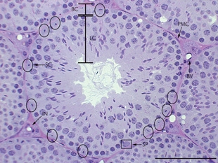
English: Junctional complexes are formed between the adjacent Sertoli cells (Sertoli-Sertoli barrier or blood-testis barrier) and this barrier separate the space within the seminiferous tubule into basal and luminal compartments. A layer of peritubular myoid cells surround the tubules. The nuclei of Sertoli cells are circled and nuclei of spermatogonia are enclosed by a rectangle. Abbreviations: BC- basal compartment; LC- luminal compartment; IN- interstitium; BV- blood vessel; SC- Sertoli cell; SP- spermatogonia; PMC- peritubular myoid cell. Bar represents 100 μm. |
| Date | Published July 14, 2008. |
| Source |
[1] Direct
|
| Author | Zhou, Q. and Griswold, M.D., Regulation of spermatogonia (July 14, 2008), StemBook, ed. The Stem Cell Research Community, StemBook, doi/10.3824/stembook.1.7.1, http://www.stembook.org. |
| Permission (Reusing this file) |
This file is licensed under the Creative Commons Attribution 3.0 Unported license.
|
File history
Click on a date/time to view the file as it appeared at that time.
| Date/Time | Thumbnail | Dimensions | User | Comment | |
|---|---|---|---|---|---|
| current | 19:04, 5 April 2013 | | 310 × 233 (69 KB) | Smallbot (talk | contribs) | Uploading CC-BY images from the the StemBook http://www.stembook.org/ 154/173 |
You cannot overwrite this file.
File usage on Commons
There are no pages that use this file.
File usage on other wikis
The following other wikis use this file:
- Usage on en.wikipedia.org
- Usage on pt.wikipedia.org