File:Echinococcus vogeli2.jpg
(Redirected from File:Ev2.jpg)
Size of this preview: 800 × 526 pixels. Other resolutions: 320 × 210 pixels | 640 × 421 pixels | 1,024 × 673 pixels | 1,280 × 841 pixels | 3,059 × 2,010 pixels.
Original file (3,059 × 2,010 pixels, file size: 1.65 MB, MIME type: image/jpeg)
Captions
Captions
Add a one-line explanation of what this file represents
| Description |
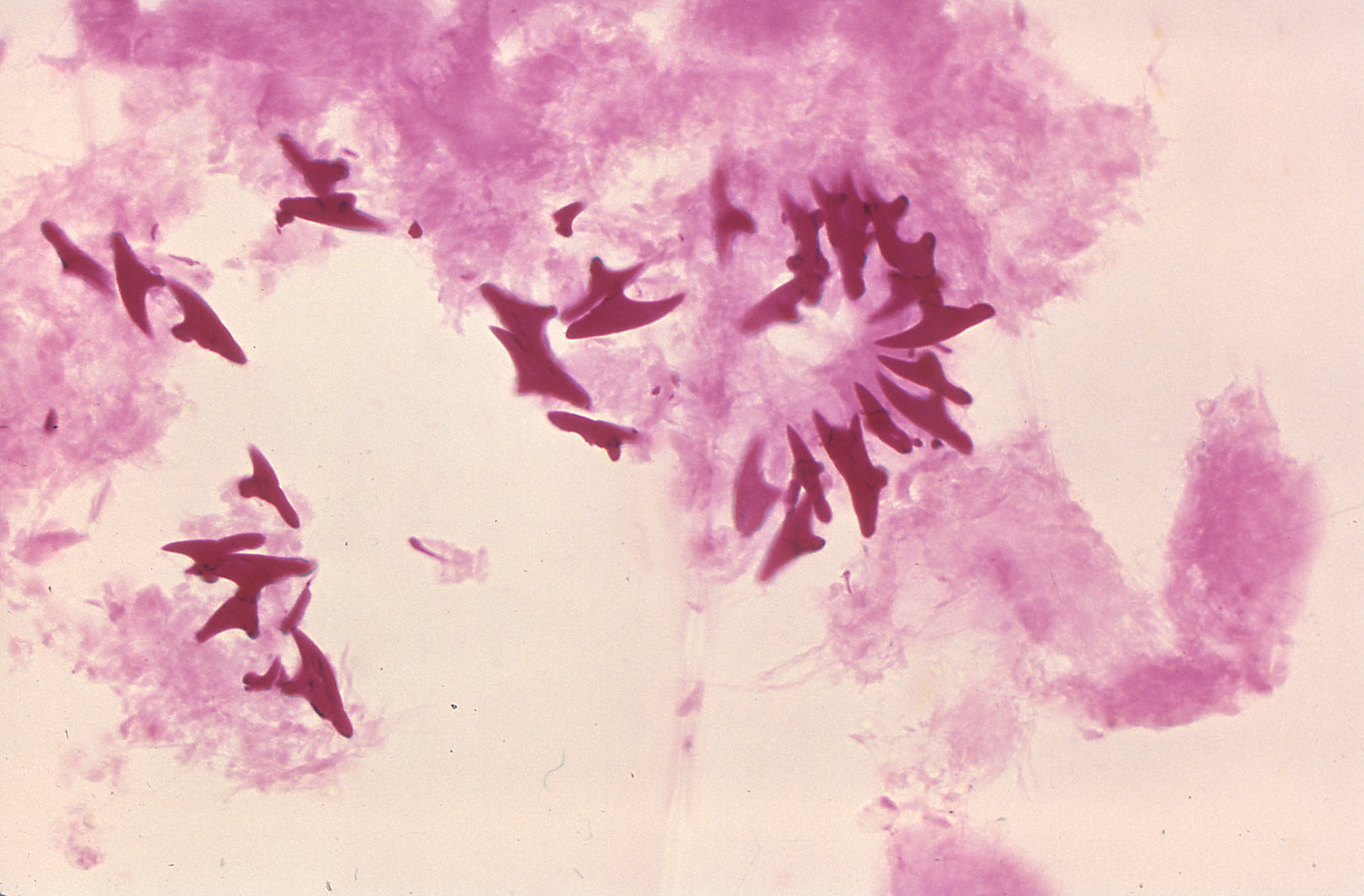
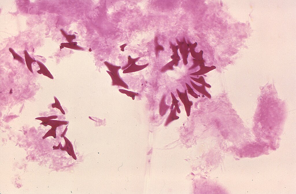
English: This photomicrograph of a tissue sample harvested from a cyst, which was harbored inside a gorilla, revealed the presence of these protoscolex hooks of a microscopic Echinococcus vogeli tapeworm. The larval stage of this microscopic tapeworm is one of the causative agents of alveolar hydatid disease (AHD), an infection in humans that causes parasitic tumors to form, mainly in the liver, but can also appear in other organs as well. |
||
| Date | |||
| Source |
|
||
| Author | CDC/Dr. Peter Schantz |
This image is a work of the Centers for Disease Control and Prevention, part of the United States Department of Health and Human Services, taken or made as part of an employee's official duties. As a work of the U.S. federal government, the image is in the public domain.
|
File history
Click on a date/time to view the file as it appeared at that time.
| Date/Time | Thumbnail | Dimensions | User | Comment | |
|---|---|---|---|---|---|
| current | 07:08, 4 August 2020 | | 3,059 × 2,010 (1.65 MB) | TommyG (talk | contribs) | Larger version from source |
| 05:19, 4 August 2020 |  | 700 × 477 (46 KB) | WQUlrich (talk | contribs) | Reverted to version as of 13:34, 18 November 2007 (UTC) | |
| 15:56, 12 March 2008 |  | 417 × 488 (178 KB) | EraserGirl (talk | contribs) | rmv border | |
| 08:31, 27 February 2008 |  | 640 × 480 (63 KB) | Musaraigne (talk | contribs) | {{Information |Description=Eugène Viala dans son atelier |Source=travail personnel |Date=2007 |Author= Musaraigne |Permission= |other_versions= }} | |
| 13:34, 18 November 2007 |  | 700 × 477 (46 KB) | Filip em (talk | contribs) | {{Information |Description=This is a photomicrograph of protoscolex hooks of Echinococcus vogeli taken from a cyst within a gorilla. The larval stage of the microscopic tapeworm Echinococcus vogeli is one of the causative agents of Alveolar Hydatid Disea |
You cannot overwrite this file.
File usage on Commons
The following page uses this file:
- File:Ev2.jpg (file redirect)