File:E01333-20F3.large.jpg
Jump to navigation
Jump to search

Size of this preview: 448 × 600 pixels. Other resolutions: 179 × 240 pixels | 358 × 480 pixels | 956 × 1,280 pixels.
Original file (956 × 1,280 pixels, file size: 277 KB, MIME type: image/jpeg)
Captions
Captions
Add a one-line explanation of what this file represents
Summary
[edit]
| Description |
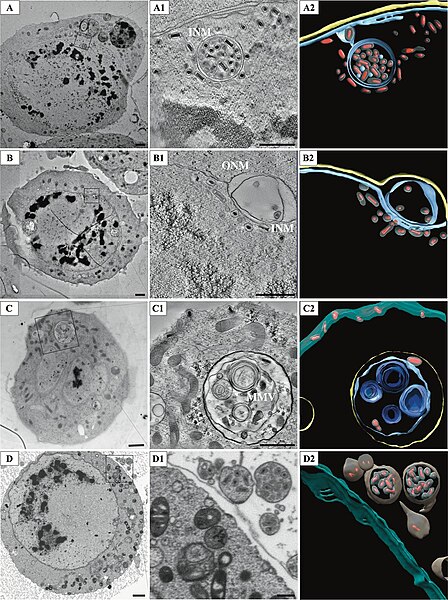
English: Oryctes rhinoceros nudivirus (OrNV) Virion trafficking inside the host cell. (A) Micrograph of an OrNV-infected cell showing virions encapsulated into a vesicle inside the nucleus. Scale bar, 1 μm. (A1) A 11-nm slice through the tomographic reconstruction indicated in inset showing virions enclosed in a double membrane vesicle. Scale bar, 500 nm. (A2) Segmentation of the tomogram. (B) Micrograph displaying expanded NE. Scale bar, 1 μm. (B1) A 11-nm slice through the tomographic reconstruction indicated in inset shows virions inside an expanded NE lumen. Scale bar, 500 nm. (B2) Segmentation of B1 highlights vesicle wrapped virions inside the NE lumen. (C) Micrograph shows cytoplasm completely traversing the nucleus. Scale bar, 1 μm. (C1) A 11-nm slice through the tomographic reconstruction of the area indicated in inset displaying virions encapsulated into multimembrane vesicles (MMVs) in the cytoplasm. Scale bar, 500 nm. (C2) Segmentation showing virions inside complex MMVs, and vesicle-free virions close to the cellular membrane. (D) An infected cell showing vesicles being expelled into the extracellular space. Scale bar, 1 μm. (D1) A slice through the tomographic reconstruction of the area indicated in inset showing vesicles containing fully enveloped virions. Scale bar, 500 nm. (D2) Segmentation showing virions within vesicles. |
| Date | |
| Source |
Fig. 3 at https://mbio.asm.org/content/11/4/e01333-20/figures-only Visualizing Nudivirus Assembly and Egress. In: ASM mBio 11:e01333-20. PMID 32788378; doi:10.1128/mBio.01333-20 |
| Author | Sailakshmi Velamoor, Allan Mitchell, Bruno M. Humbel, WonMo Kim, Charlotte Pushparajan, Gabriel Visnovsky, Laura N. Burga, Mihnea Bostina; Anne Moscona (Editor) |
| Other versions |
      |
Licensing
[edit]
This file is licensed under the Creative Commons Attribution-Share Alike 4.0 International license.
- You are free:
- to share – to copy, distribute and transmit the work
- to remix – to adapt the work
- Under the following conditions:
- attribution – You must give appropriate credit, provide a link to the license, and indicate if changes were made. You may do so in any reasonable manner, but not in any way that suggests the licensor endorses you or your use.
- share alike – If you remix, transform, or build upon the material, you must distribute your contributions under the same or compatible license as the original.
File history
Click on a date/time to view the file as it appeared at that time.
| Date/Time | Thumbnail | Dimensions | User | Comment | |
|---|---|---|---|---|---|
| current | 09:12, 21 April 2021 | | 956 × 1,280 (277 KB) | Ernsts (talk | contribs) | Uploaded a work by Sailakshmi Velamoor, Allan Mitchell, Bruno M. Humbel, WonMo Kim, Charlotte Pushparajan, Gabriel Visnovsky, Laura N. Burga, Mihnea Bostina; Anne Moscona (Editor) from https://mbio.asm.org/content/11/4/e01333-20/figures-only Visualizing Nudivirus Assembly and Egress. In: ASM mBio 11:e01333-20. PMID 32788378; doi:10.1128/mBio.01333-20 50px|class=noviewer with UploadWizard |
You cannot overwrite this file.
File usage on Commons
The following 6 pages use this file: