File:Drosophila embryo musculoskeletal system.jpg
Jump to navigation
Jump to search

Size of this preview: 800 × 417 pixels. Other resolutions: 320 × 167 pixels | 640 × 333 pixels | 1,024 × 533 pixels | 1,490 × 776 pixels.
Original file (1,490 × 776 pixels, file size: 375 KB, MIME type: image/jpeg)
Captions
Captions
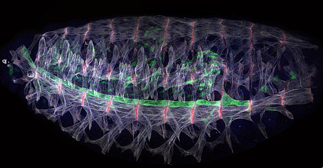
This image shows the musculoskeletal system of the stage 16 Drosophila embryo
Summary[edit]
| Description |
English: Nascent myotubes undergo a dramatic morphological transformation during myogenesis, in which the myotubes elongate over several cell diameters and are directed to the correct muscle attachment sites. This image shows the musculoskeletal system of the stage 16 Drosophila embryo. 30 distinct muscles were stained with anti-Tm (white) and VL1 muscle is specifically identified by the Gal4 system (5053-Gal4>UAS-GFP). |
| Date | |
| Source | Own work |
| Author | Shuo Yang |
Licensing[edit]
I, the copyright holder of this work, hereby publish it under the following license:
This file is licensed under the Creative Commons Attribution 4.0 International license.
- You are free:
- to share – to copy, distribute and transmit the work
- to remix – to adapt the work
- Under the following conditions:
- attribution – You must give appropriate credit, provide a link to the license, and indicate if changes were made. You may do so in any reasonable manner, but not in any way that suggests the licensor endorses you or your use.
| This file was uploaded as part of Wiki Science Competition 2021. |
File history
Click on a date/time to view the file as it appeared at that time.
| Date/Time | Thumbnail | Dimensions | User | Comment | |
|---|---|---|---|---|---|
| current | 05:57, 28 November 2021 | | 1,490 × 776 (375 KB) | Lmys2356 (talk | contribs) | Uploaded own work with UploadWizard |
You cannot overwrite this file.
File usage on Commons
The following 2 pages use this file:
File usage on other wikis
The following other wikis use this file:
- Usage on en.wikipedia.org