File:Asterostegus tuberculatus (10.5852-ejt.2014.76) Figure 8.jpg
Jump to navigation
Jump to search

Size of this preview: 510 × 599 pixels. Other resolutions: 204 × 240 pixels | 409 × 480 pixels | 654 × 768 pixels | 872 × 1,024 pixels | 2,278 × 2,675 pixels.
Original file (2,278 × 2,675 pixels, file size: 2.34 MB, MIME type: image/jpeg)
Captions
Captions
Add a one-line explanation of what this file represents
Summary
[edit]_Figure_8.jpg&action=edit§ion=1)
| Description |
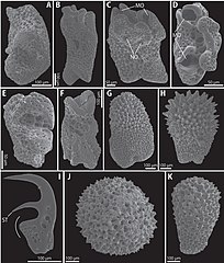
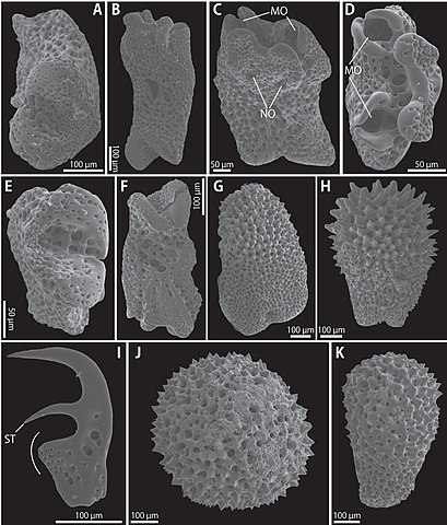
English: Fig. 8. Asterostegus tuberculatus Mortensen, 1933 (SMNH 123461), SEM photographs. A-C. Lateral arm plates from middle portion of arm: internal view (A), oral view (B) and external view (C). D-F. Late-ral arm plates from distal portion of arm: external view (D), internal view (E) and oral view (F). G-I. Arm spines from proximal portion (G), middle portion (H) and distal portion, an arc indicates lamina (I). J-K. Tubercles on proximal portion of arm: external view (J) and lateral view (K). Abbreviations: MO = muscle opening, NO = nerve opening, ST = secondary tooth. |
| Date | |
| Source | https://dx.doi.org/10.5852%2Fejt.2014.76 |
| Author | Okanishi, M.; Fujita, T. 2014: A taxonomic review of the genus Asterostegus (Echinodermata: Ophiuroidea), with the description of a new species. European journal of taxonomy, (76) DOI: 10.5852/ejt.2014.76 |
| Permission (Reusing this file) |
This file is licensed under the Creative Commons Attribution 3.0 Unported license.
|
File history
Click on a date/time to view the file as it appeared at that time.
| Date/Time | Thumbnail | Dimensions | User | Comment | |
|---|---|---|---|---|---|
| current | 11:34, 10 October 2020 | | 2,278 × 2,675 (2.34 MB) | Christian Ferrer (talk | contribs) | {{Information | description = {{en|1=Fig. 8. ''Asterostegus tuberculatus'' Mortensen, 1933 (SMNH 123461), SEM photographs. A-C. Lateral arm plates from middle portion of arm: internal view (A), oral view (B) and external view (C). D-F. Late-ral arm plates from distal portion of arm: external view (D), internal view (E) and oral view (F). G-I. Arm spines from proximal portion (G), middle portion (H) and distal portion, an arc indicates lamina (I). J-K. Tubercles on proximal portion of arm: ex... |
You cannot overwrite this file.
File usage on Commons
There are no pages that use this file.
_Figure_8.jpg&oldid=805646043)