File:Armadillidium vulgare (10.1590-2358-2936e2020041) Figure 2.jpg
Jump to navigation
Jump to search

Size of this preview: 485 × 599 pixels. Other resolutions: 194 × 240 pixels | 389 × 480 pixels | 1,000 × 1,235 pixels.
Original file (1,000 × 1,235 pixels, file size: 689 KB, MIME type: image/jpeg)
Captions
Captions
Add a one-line explanation of what this file represents
Summary[edit]
_Figure_2.jpg&action=edit§ion=1)
| Description |
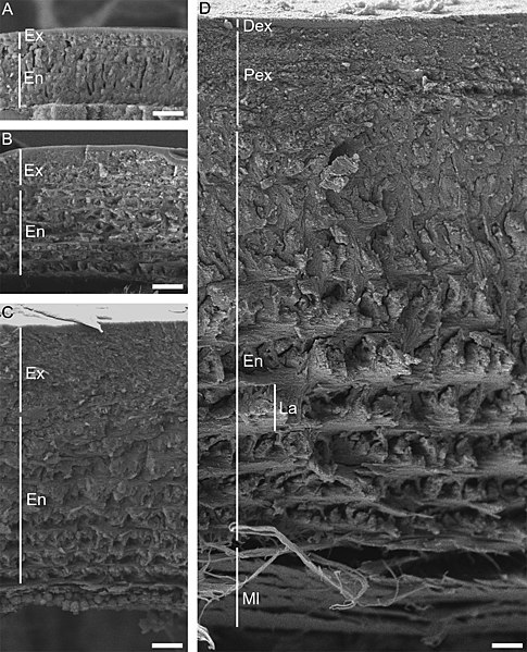
English: Figure 2. Scanning electron micrographs of sagittally fractured anterior tergites of the pereon from Armadillidium vulgare individuals of different sizes, shown to scale. A, Manca larva; 1.5 mm body length. B, 2.2 mm body length. C: 4.5 mm body length. D, 11 mm body length. Scale bars = 2 µm. Dex: distal exocuticle; En: endocuticle; Ex: exocuticle; La: lamella in the endocuticle; Ml: membranous layer; Pex: proximal exocuticle. |
| Date | |
| Source | https://doi.org/10.1590/2358-2936e2020041 |
| Author | [Photographs by Ana Sterle] Vittori, M., Vodnik, K. and Blejec, A. 2020. Changes in cuticle structure during growth in two terrestrial isopods (Crustacea: Isopoda: Oniscidea). Nauplius. 28: e2020041 |
| Permission (Reusing this file) |
This file is licensed under the Creative Commons Attribution 4.0 International license.
|
File history
Click on a date/time to view the file as it appeared at that time.
| Date/Time | Thumbnail | Dimensions | User | Comment | |
|---|---|---|---|---|---|
| current | 18:48, 28 May 2021 | | 1,000 × 1,235 (689 KB) | Christian Ferrer (talk | contribs) | {{Information | description = {{en|1=Figure 2. Scanning electron micrographs of sagittally fractured anterior tergites of the pereon from ''Armadillidium vulgare'' individuals of different sizes, shown to scale. A, Manca larva; 1.5 mm body length. B, 2.2 mm body length. C: 4.5 mm body length. D, 11 mm body length. Scale bars = 2 µm. Dex: distal exocuticle; En: endocuticle; Ex: exocuticle; La: lamella in the endocuticle; Ml: membranous layer; Pex: proximal exocuticle.}} | date = 2020-10-28... |
You cannot overwrite this file.
File usage on Commons
There are no pages that use this file.
_Figure_2.jpg&oldid=801615326)