File:Apophyllite 100.png
Jump to navigation
Jump to search

Size of this preview: 607 × 600 pixels. Other resolutions: 243 × 240 pixels | 486 × 480 pixels | 777 × 768 pixels | 1,149 × 1,135 pixels.
Original file (1,149 × 1,135 pixels, file size: 168 KB, MIME type: image/png)
Captions
Captions
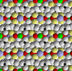
Structure of apophyllite viewed in the {100} direction, parallel to layering
Summary[edit]
| Description |
English: This image depicts the structure of apophyllite viewed along the {100} axis, parallel to the layering. T layers (composed of silica tetrahedra) are bonded together by hydroxyl, fluorine, calcium, and potassium ions. White spheres are oxygen; small black spheres are silicon; yellow spheres are hydroxyl; red spheres are calcium; blue spheres are potassium; and green spheres are fluorine. Structure from Ståhl, Kenny (1 January 1993). "A neutron powder diffraction study of partially dehydrated fluorapophyllite, KCa4Si8O20F.6.9H2O". European Journal of Mineralogy. 5 (5): 845–850. doi:10.1127/ejm/5/5/0845. |
| Date | |
| Source | Own work |
| Author | Kent G. Budge |
Licensing[edit]
I, the copyright holder of this work, hereby publish it under the following license:
| This file is made available under the Creative Commons CC0 1.0 Universal Public Domain Dedication. | |
| The person who associated a work with this deed has dedicated the work to the public domain by waiving all of their rights to the work worldwide under copyright law, including all related and neighboring rights, to the extent allowed by law. You can copy, modify, distribute and perform the work, even for commercial purposes, all without asking permission.
|
File history
Click on a date/time to view the file as it appeared at that time.
| Date/Time | Thumbnail | Dimensions | User | Comment | |
|---|---|---|---|---|---|
| current | 16:20, 20 April 2021 | | 1,149 × 1,135 (168 KB) | Kent G. Budge (talk | contribs) | Uploaded own work with UploadWizard |
You cannot overwrite this file.
File usage on Commons
There are no pages that use this file.
File usage on other wikis
The following other wikis use this file:
- Usage on en.wikipedia.org
- Usage on uz.wikipedia.org