File:Anatomy of an egg c-m.svg
Jump to navigation
Jump to search

Size of this PNG preview of this SVG file: 483 × 599 pixels. Other resolutions: 193 × 240 pixels | 387 × 480 pixels | 619 × 768 pixels | 826 × 1,024 pixels | 1,651 × 2,048 pixels | 566 × 702 pixels.
Original file (SVG file, nominally 566 × 702 pixels, file size: 43 KB)
Captions
Captions
Add a one-line explanation of what this file represents
Summary[edit]
Modyfied Version of Anatomy of an egg.svg
| Description |
Deutsch:
Schematischer Längsschnitt eines Hühnereis:
Čeština:
Stavba ptačího vejce:
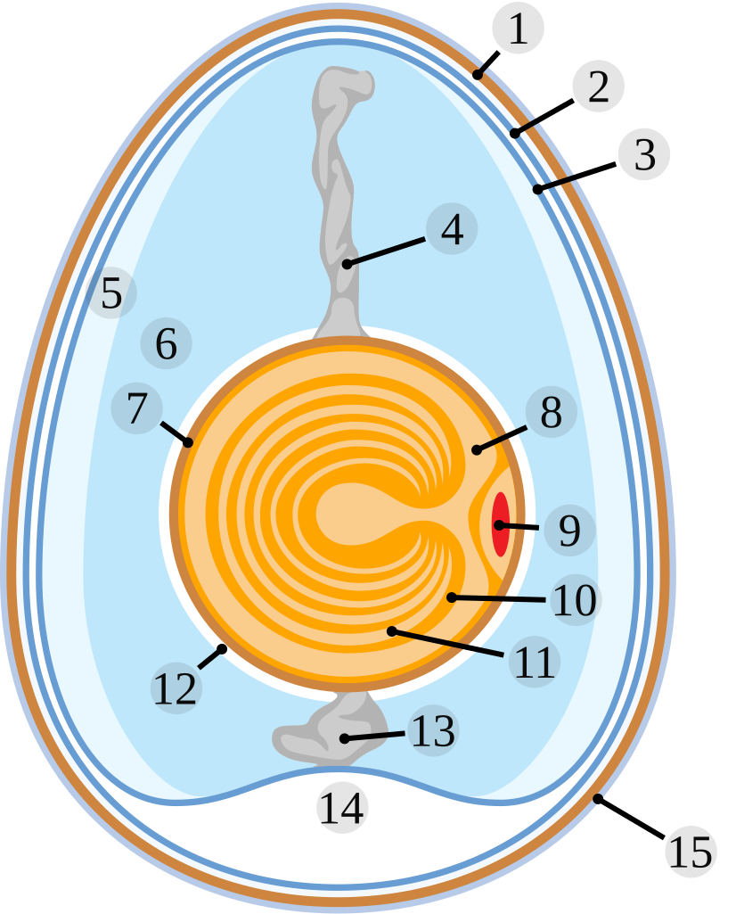
English:
Schematic of a chicken egg:
Italiano:
Morfologia dell'uovo di pollo:
Nederlands: Schematische voorstelling van een kippenei:
|
| Date | |
| Source | graphic created by Horst Frank, SVG version by -xfi- |
| Author | Horst Frank, SVG code -xfi- |
| Permission (Reusing this file) |
Horst Frank released it under GFDL |
| Other versions | Ei1.jpg ; corrected : Anatomy of an amiotic egg.svg |
| SVG development | This file uses translateable embedded text. |
Licensing[edit]

|
Permission is granted to copy, distribute and/or modify this document under the terms of the GNU Free Documentation License, Version 1.2 or any later version published by the Free Software Foundation; with no Invariant Sections, no Front-Cover Texts, and no Back-Cover Texts. A copy of the license is included in the section entitled GNU Free Documentation License. |
| This file is licensed under the Creative Commons Attribution-Share Alike 3.0 Unported license. | ||
| ||
| This licensing tag was added to this file as part of the GFDL licensing update. |
File history
Click on a date/time to view the file as it appeared at that time.
| Date/Time | Thumbnail | Dimensions | User | Comment | |
|---|---|---|---|---|---|
| current | 13:19, 13 January 2007 | | 566 × 702 (43 KB) | C-M (talk | contribs) | |
| 13:10, 13 January 2007 |  | 566 × 702 (40 KB) | C-M (talk | contribs) | ||
| 11:50, 13 January 2007 |  | 566 × 702 (27 KB) | C-M (talk | contribs) | ||
| 11:42, 13 January 2007 |  | 566 × 702 (27 KB) | C-M (talk | contribs) | Modyfied Version of Image:Anatomy of an egg.svg {{Information| |Description = {{de|<br />Schematischer Längsschnitt eines Hühnereis: #Kalkschale #Schalenhaut #Schalenhaut #Chalaza (Hagelschnur) #äußeres Eiklar (dünnflüssig) #mittleres Eiklar (d |
You cannot overwrite this file.
File usage on Commons
The following 5 pages use this file:
File usage on other wikis
The following other wikis use this file:
- Usage on ca.wikipedia.org
- Usage on de.wikipedia.org
- Usage on es.wikipedia.org
- Usage on eu.wikipedia.org
- Usage on gl.wikipedia.org
- Usage on it.wikipedia.org
- Usage on ko.wikipedia.org
- Usage on nl.wikipedia.org
- Usage on nl.wiktionary.org
- Usage on no.wikipedia.org
- Usage on pl.wikipedia.org
- Usage on pl.wiktionary.org
- Usage on pt.wikipedia.org
- Usage on ru.wikipedia.org
- Usage on uk.wikipedia.org