File:An example of a lysibody.jpg
Jump to navigation
Jump to search
Size of this preview: 800 × 235 pixels. Other resolutions: 320 × 94 pixels | 640 × 188 pixels | 1,024 × 300 pixels | 3,119 × 915 pixels.
Original file (3,119 × 915 pixels, file size: 84 KB, MIME type: image/jpeg)
Captions
Captions
Add a one-line explanation of what this file represents
Summary[edit]
| Description |
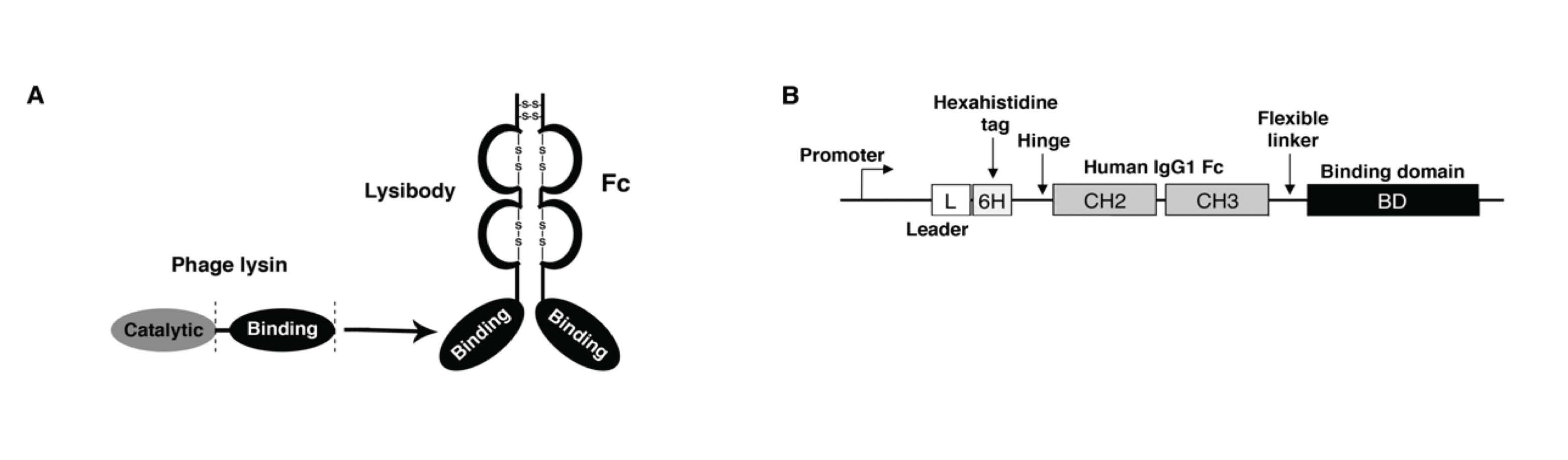
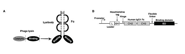
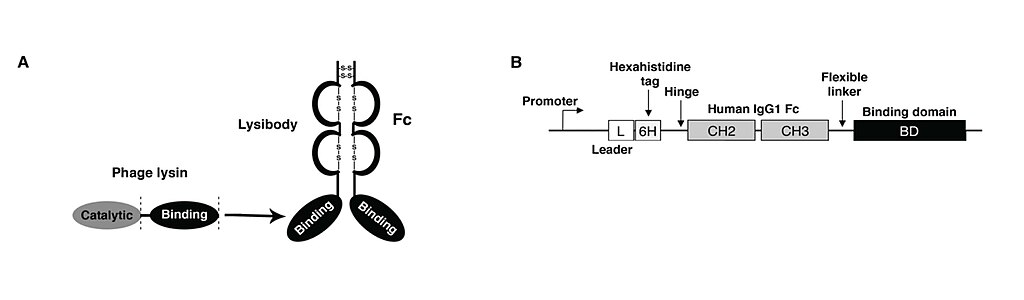
English: A. The binding domain from a phage lysin is linked to the C-terminal end of the human IgG1 Fc. The cysteine in the hinge region of the IgG1 will form S-S bonds to produce a stable homodimer.
B. How the lysibody is constructed: A promotor followed by the leader sequence (L) and a his tag (6H) for ease in purification. This is followed by the hinge region of human IgG 1 and human IgG1 Fc. After a short linker, the binding domain of a phage lysin in linked at the C-terminal end of the lysibody molecule. |
| Date | |
| Source | Own work |
| Author | Vaf222 |
Licensing[edit]
I, the copyright holder of this work, hereby publish it under the following license:
This file is licensed under the Creative Commons Attribution-Share Alike 4.0 International license.
- You are free:
- to share – to copy, distribute and transmit the work
- to remix – to adapt the work
- Under the following conditions:
- attribution – You must give appropriate credit, provide a link to the license, and indicate if changes were made. You may do so in any reasonable manner, but not in any way that suggests the licensor endorses you or your use.
- share alike – If you remix, transform, or build upon the material, you must distribute your contributions under the same or compatible license as the original.
File history
Click on a date/time to view the file as it appeared at that time.
| Date/Time | Thumbnail | Dimensions | User | Comment | |
|---|---|---|---|---|---|
| current | 19:50, 21 January 2019 | 3,119 × 915 (84 KB) | Vaf222 (talk | contribs) | Cross-wiki upload from en.wikipedia.org |
You cannot overwrite this file.
File usage on Commons
The following page uses this file:
- File:An example of a lysibody .jpg (file redirect)
File usage on other wikis
The following other wikis use this file:
- Usage on en.wikipedia.org