File:A What is the differentiation potential of cells in the blastema?.jpg
From Wikimedia Commons, the free media repository
Jump to navigation
Jump to search
A_What_is_the_differentiation_potential_of_cells_in_the_blastema?.jpg (531 × 600 pixels, file size: 93 KB, MIME type: image/jpeg)
File information
Structured data
Captions
Captions
Add a one-line explanation of what this file represents
| DescriptionA What is the differentiation potential of cells in the blastema?.jpg |
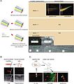
English: The blastema may be composed of 1) cells that are restricted to give rise to the same tissues from which they were derived, 2) cells that are multipotent and give rise different tissue types, or 3) a complex mix of cells with a variety of origins and potentials. In the larval tail of urodeles, fluorescently tagged glial cells of the spinal cord proliferate during regeneration and can give rise to tissue outside of the spinal cord. Data currently indicates that at least in larval tissues, the urodele blastema contains a complex mix of lineage-restricted and multipotent cells. However, very little is known about the potential of blastema cells in adult urodeles. In anuran amphibians, GFP+ tissue grafts into GFP- hosts illustrate that cells of the regenerating spinal cord and notochord are derived from cells of those very same tissues. Therefore, the progenitor cells during anuran tadpole regeneration appear to be restricted in their potential. B Injection of dye into multinucleated muscle fibers prior to amputation illustrates that once clipped, some these fibers can fragment into proliferating mononuclear cells that contribute to the blastema. The long-term fate of these cells is still under active investigation. C (left) Muscle fibers in adult newts contain satellite-like cells that express Pax7 (green) and are separated from the rest of the cell by a basement membrane (red). (middle, right) When these cells are isolated, cultured, tagged (red) and implanted into regenerating newt limbs, they contribute to the blastema and give rise to unrelated tissue types including cartilage (green) and epidermis (green). Images are adapted from: (1) Echeverri, K., and Tanaka, E. M. (2002). Ectoderm to mesoderm lineage switching during axolotl tail regeneration. Science 298, 1993–1996. Reprinted with permission from AAAS. (2) Gargioli, C., and Slack, J. M. (2004). Cell lineage tracing during Xenopus tail regeneration. Development 131, 2669–2679. Reproduced with permission of the Company of Biologists. (3) Reprinted from Cell, 113, Tanaka, E. M., Regeneration: if they can do it, why can't we?, 559–562, 2003, with permission from Elsevier. (4) Echeverri, K., Clarke, J. D., and Tanaka, E. M. (2001). In vivo imaging indicates muscle fiber dedifferentiation is a major contributor to the regenerating tail blastema. Dev Biol 236, 151–164. (5) Kumar et al., 2004, PLOS Biology, (6) © Morrison et al., 2006. Originally published in The Journal of Cell Biology. doi:10.1083/jcb.200509011. |
| Date | Published December 31, 2008. |
| Source |
[1] Direct
|
| Author | Gurley, K.A. and Sánchez Alvarado, A., Stem cells in animal models of regeneration (December 31, 2008), StemBook, ed. The Stem Cell Research Community, StemBook, doi/10.3824/stembook.1.32.1, http://www.stembook.org. |
| Permission (Reusing this file) |
This file is licensed under the Creative Commons Attribution 3.0 Unported license.
|
File history
Click on a date/time to view the file as it appeared at that time.
| Date/Time | Thumbnail | Dimensions | User | Comment | |
|---|---|---|---|---|---|
| current | 18:49, 5 April 2013 |  | 531 × 600 (93 KB) | Smallbot (talk | contribs) | Uploading CC-BY images from the the StemBook http://www.stembook.org/ 64/173 |
You cannot overwrite this file.
File usage on Commons
There are no pages that use this file.
Structured data
Items portrayed in this file
depicts
image/jpeg
5a9f6681f8d14f9e5ebb229c5c6c02d93949ab97
95,601 byte
600 pixel
531 pixel
Hidden categories:
