File:Waginella ebonita (10.3389-fmars.2021.616001) Figure 8.jpg
Jump to navigation
Jump to search
Size of this preview: 623 × 600 pixels. Other resolutions: 249 × 240 pixels | 498 × 480 pixels | 797 × 768 pixels | 1,063 × 1,024 pixels | 1,869 × 1,800 pixels.
Original file (1,869 × 1,800 pixels, file size: 1.51 MB, MIME type: image/jpeg)
Captions
Captions
Add a one-line explanation of what this file represents
Summary
[edit]_Figure_8.jpg&action=edit§ion=1)
| Description |
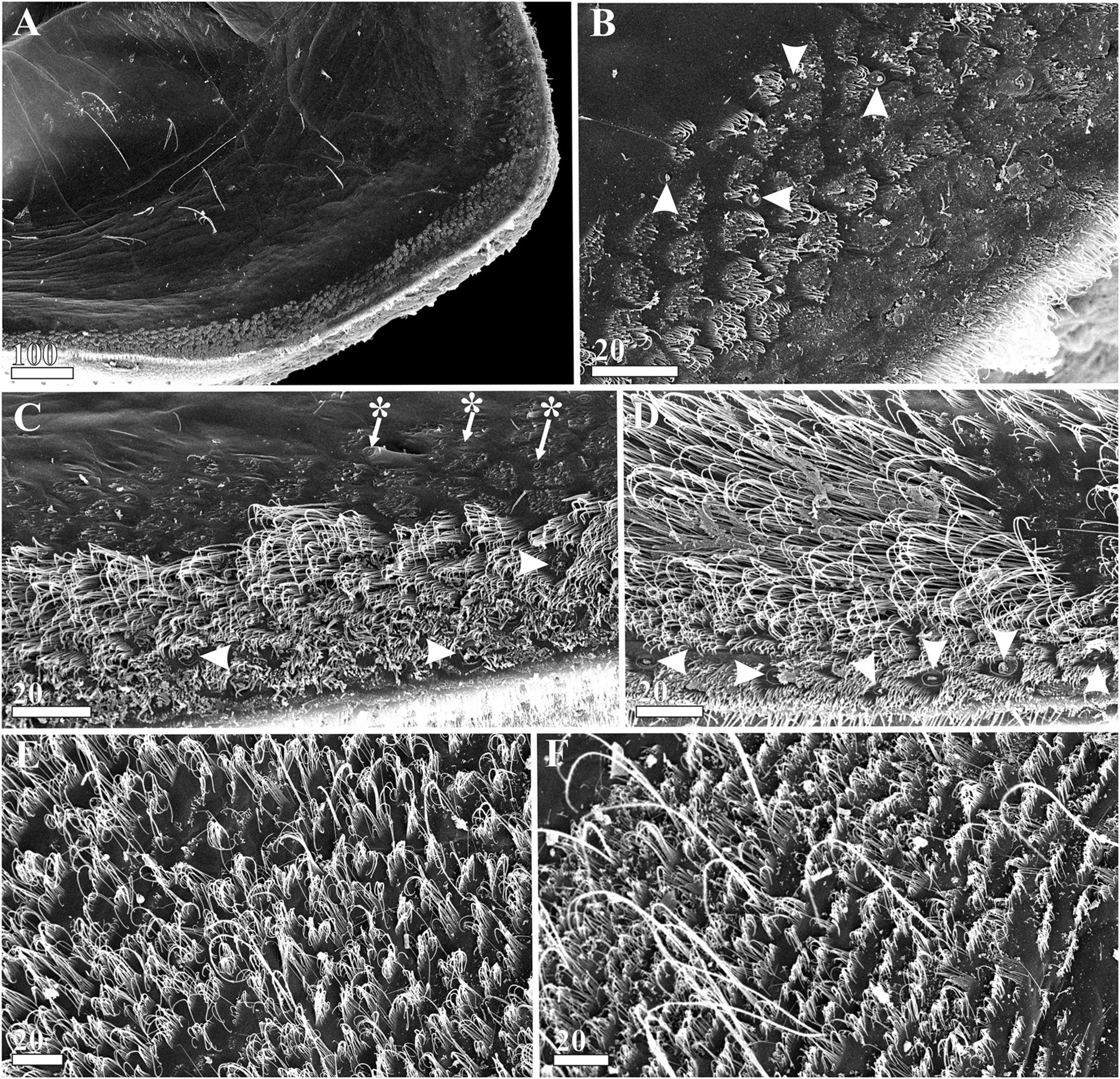
English: Figure 8. Waginella ebonita sp. nov., female, holotype. Carapace mantle structures (SEM). (A) Surface of mantle, anterioventral part. (B) Ornamentation of mantle surface at anterior margin (gland tubes indicated by arrowheads). (C) Ornamentation of mid-ventral part of mantle surface (gland tubes indicated by arrowheads, cuticular pores—by asterisks). (D) Ornamentation of posterioventral part of mantle surface (marginal gland tubes indicated by arrowheads). (E) Ornamentation of mantle surface at posterior margin. (F) Ornamentation of mantle surface at posteriodorsal margin. Scale bars in μm. |
| Date | |
| Source | Kolbasov GA, Savchenko AS, Newman WA and Chan BKK (2021) A New Species of Waginella (Crustacea: Thecostraca: Ascothoracida) Parasitic on a Stalked Crinoid From Tasman Sea, With Notes on Morphology of Related Genera. Front. Mar. Sci. 8:616001. https://doi.org/10.3389/fmars.2021.616001 |
| Author | Kolbasov GA, Savchenko AS, Newman WA and Chan BKK (2021) |
| Permission (Reusing this file) |
This file is licensed under the Creative Commons Attribution 4.0 International license.
|
File history
Click on a date/time to view the file as it appeared at that time.
| Date/Time | Thumbnail | Dimensions | User | Comment | |
|---|---|---|---|---|---|
| current | 05:44, 19 December 2022 | | 1,869 × 1,800 (1.51 MB) | Christian Ferrer (talk | contribs) | {{Information | description = {{en|1=Figure 8. ''Waginella ebonita'' sp. nov., female, holotype. Carapace mantle structures (SEM). (A) Surface of mantle, anterioventral part. (B) Ornamentation of mantle surface at anterior margin (gland tubes indicated by arrowheads). (C) Ornamentation of mid-ventral part of mantle surface (gland tubes indicated by arrowheads, cuticular pores—by asterisks). (D) Ornamentation of posterioventral part of mantle surface (marginal gland tubes indicated by arrowhe... |
You cannot overwrite this file.
File usage on Commons
There are no pages that use this file.
_Figure_8.jpg&oldid=717681013)