File:Waginella ebonita (10.3389-fmars.2021.616001) Figure 5.jpg
Jump to navigation
Jump to search

Size of this preview: 485 × 599 pixels. Other resolutions: 194 × 240 pixels | 388 × 480 pixels | 622 × 768 pixels | 829 × 1,024 pixels | 1,781 × 2,200 pixels.
Original file (1,781 × 2,200 pixels, file size: 648 KB, MIME type: image/jpeg)
Captions
Captions
Add a one-line explanation of what this file represents
Summary
[edit]_Figure_5.jpg&action=edit§ion=1)
| Description |
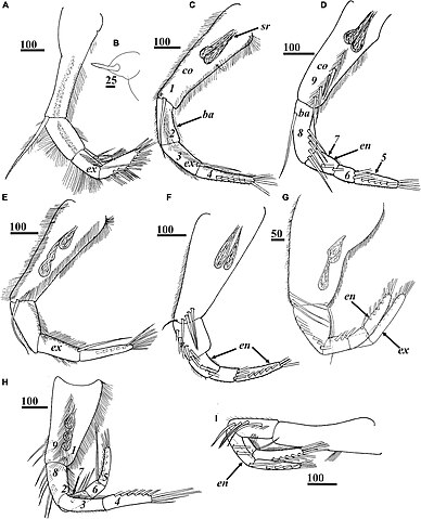
English: Figure 5. Waginella ebonita sp. nov., female, holotype. (A) Left thoracopod 1. (B) Filamentary appendage at base of thoracopod 1. (C,D) Left and right thoracopods 2. (E,F) Left and right thoracopods 3. (G) Right thoracopod 4. (H) Left thoracopod 5. (I) Right thoracopod 6. Ampuliform seminal receptacles are situated in upper outer parts of coxae of thoracopods 2–5 (C–H). Numbers indicating positions for setal counts in description (1–9) are shown for thoracopods 2 (C,D). ba, basis; co, coxa; en, endopod; ex, exopod; sr, seminal receptacles. Scale bars in μm. |
| Date | |
| Source | Kolbasov GA, Savchenko AS, Newman WA and Chan BKK (2021) A New Species of Waginella (Crustacea: Thecostraca: Ascothoracida) Parasitic on a Stalked Crinoid From Tasman Sea, With Notes on Morphology of Related Genera. Front. Mar. Sci. 8:616001. https://doi.org/10.3389/fmars.2021.616001 |
| Author | Kolbasov GA, Savchenko AS, Newman WA and Chan BKK (2021) |
| Permission (Reusing this file) |
This file is licensed under the Creative Commons Attribution 4.0 International license.
|
File history
Click on a date/time to view the file as it appeared at that time.
| Date/Time | Thumbnail | Dimensions | User | Comment | |
|---|---|---|---|---|---|
| current | 05:42, 19 December 2022 | | 1,781 × 2,200 (648 KB) | Christian Ferrer (talk | contribs) | {{Information | description = {{en|1=Figure 5. ''Waginella ebonita'' sp. nov., female, holotype. (A) Left thoracopod 1. (B) Filamentary appendage at base of thoracopod 1. (C,D) Left and right thoracopods 2. (E,F) Left and right thoracopods 3. (G) Right thoracopod 4. (H) Left thoracopod 5. (I) Right thoracopod 6. Ampuliform seminal receptacles are situated in upper outer parts of coxae of thoracopods 2–5 (C–H). Numbers indicating positions for setal counts in description (1–9) are shown for t... |
You cannot overwrite this file.
File usage on Commons
There are no pages that use this file.
_Figure_5.jpg&oldid=717678927)