File:Waginella ebonita (10.3389-fmars.2021.616001) Figure 11.jpg
Jump to navigation
Jump to search

Size of this preview: 473 × 599 pixels. Other resolutions: 189 × 240 pixels | 379 × 480 pixels | 606 × 768 pixels | 808 × 1,024 pixels | 1,737 × 2,200 pixels.
Original file (1,737 × 2,200 pixels, file size: 1.39 MB, MIME type: image/jpeg)
Captions
Captions
Add a one-line explanation of what this file represents
Summary
[edit]_Figure_11.jpg&action=edit§ion=1)
| Description |
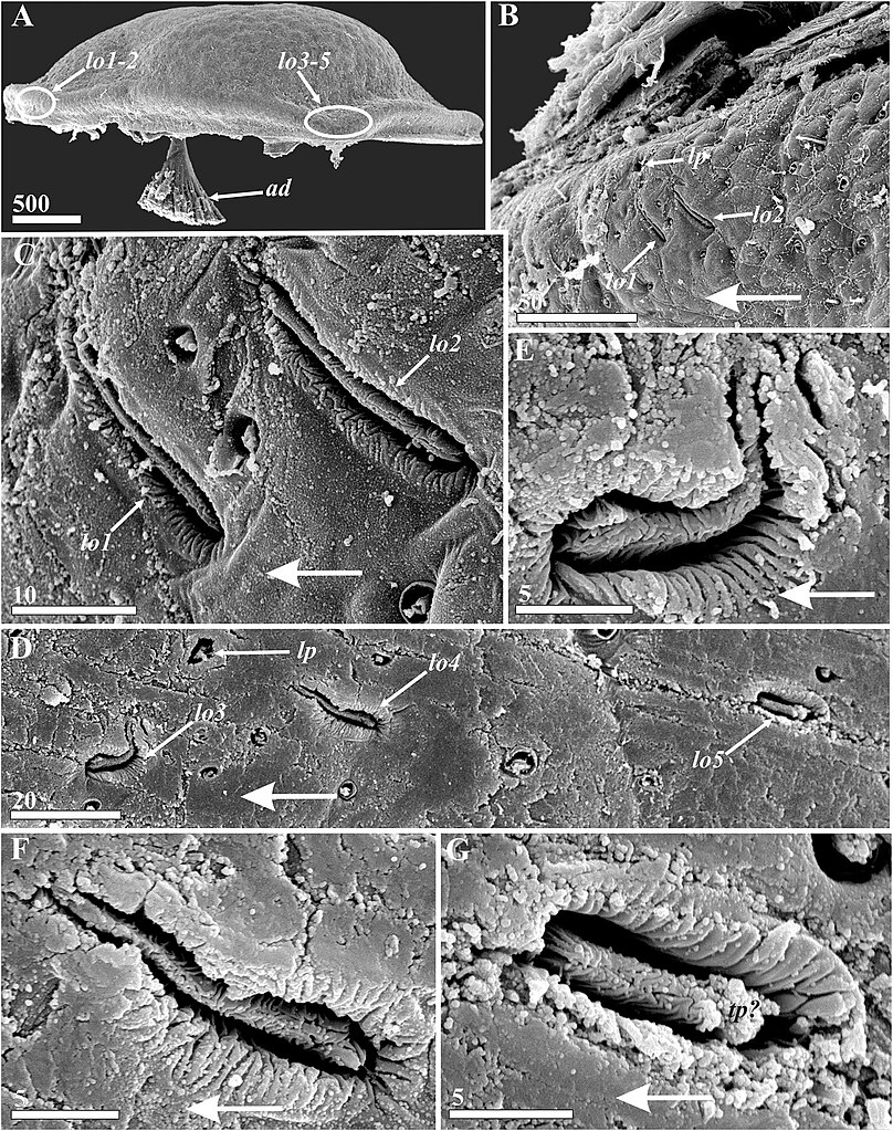
English: Figure 11. Waginella ebonita sp. nov., female, holotype. Lattice organs, with large arrows indicating anterior direction (SEM). (A) Locations of lattice organs (indicated by oval outlines) on right valve of carapace, anterior end left. (B) Anterior lattice organs (1, 2) on left carapace valve. (C) Enlarged left anterior lattice organs (1, 2). (D) Left posterior lattice organs (3–5). (E–G) Left posterior lattice organs of 3, 4, and 5 pair, respectively. ad, adductor muscle; lo1–5, lattice organs; lp, large pore associated with lattice organs; tp?, probable terminal pore of lattice organ. Scale bars in μm. |
| Date | |
| Source | Kolbasov GA, Savchenko AS, Newman WA and Chan BKK (2021) A New Species of Waginella (Crustacea: Thecostraca: Ascothoracida) Parasitic on a Stalked Crinoid From Tasman Sea, With Notes on Morphology of Related Genera. Front. Mar. Sci. 8:616001. https://doi.org/10.3389/fmars.2021.616001 |
| Author | Kolbasov GA, Savchenko AS, Newman WA and Chan BKK (2021) |
| Permission (Reusing this file) |
This file is licensed under the Creative Commons Attribution 4.0 International license.
|
File history
Click on a date/time to view the file as it appeared at that time.
| Date/Time | Thumbnail | Dimensions | User | Comment | |
|---|---|---|---|---|---|
| current | 05:46, 19 December 2022 | | 1,737 × 2,200 (1.39 MB) | Christian Ferrer (talk | contribs) | {{Information | description = {{en|1=Figure 11. ''Waginella ebonita'' sp. nov., female, holotype. Lattice organs, with large arrows indicating anterior direction (SEM). (A) Locations of lattice organs (indicated by oval outlines) on right valve of carapace, anterior end left. (B) Anterior lattice organs (1, 2) on left carapace valve. (C) Enlarged left anterior lattice organs (1, 2). (D) Left posterior lattice organs (3–5). (E–G) Left posterior lattice organs of 3, 4, and 5 pair, respectively... |
You cannot overwrite this file.
File usage on Commons
There are no pages that use this file.
_Figure_11.jpg&oldid=717920637)