File:Variant Creutzfeldt-Jakob disease (vCJD), H&E.jpg
Jump to navigation
Jump to search
No higher resolution available.
Variant_Creutzfeldt-Jakob_disease_(vCJD),_H&E.jpg (700 × 554 pixels, file size: 80 KB, MIME type: image/jpeg)
Captions
Captions
Add a one-line explanation of what this file represents
Summary
[edit],_H%26E.jpg&action=edit§ion=1)
| Description |
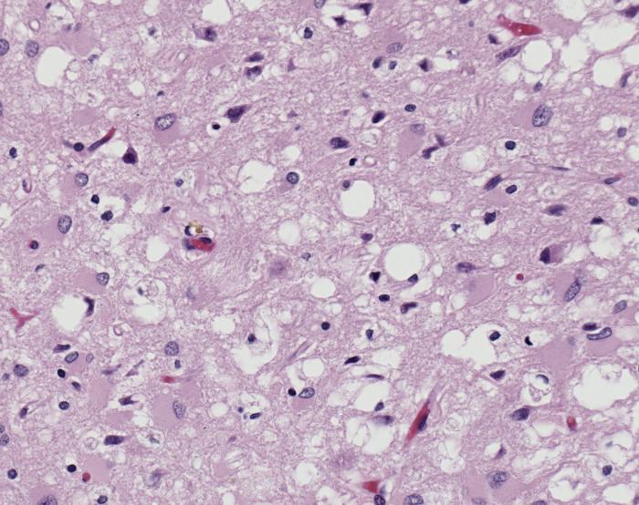
ID#: 10131 Magnified 100X, and stained with H&E (hematoxylin and eosin) staining technique, this light photomicrograph of brain tissue reveals the presence of prominent spongiotic changes in the cortex, and loss of neurons in a case of variant Creutzfeldt-Jakob disease (vCJD). Variant Creutzfeldt-Jakob disease (vCJD) is a prion disease that was first described in 1996 in the United Kingdom. There is now strong scientific evidence that the agent responsible for the outbreak of prion disease in cows, bovine spongiform encephalopathy (BSE or 'mad cow' disease), is the same agent responsible for the outbreak of vCJD in humans. Both disorders are invariably fatal brain diseases with unusually long incubation periods measured in years, and are caused by an unconventional transmissible agent called a prion. vCJD is not the same disease as classic CJD. It has different clinical and pathologic characteristics from classic CJD. Each disease also has a particular genetic profile of the prion protein gene. |
| Source | Public Health Image Library (PHIL) ID#: 10131 |
| Author |
Content Providers(s): CDC/ Teresa Hammett Photo Credit: Sherif Zaki; MD; PhD; Wun-Ju Shieh; MD; PhD; MPH |
| Permission (Reusing this file) |
Copyright Restrictions: None - This image is in the public domain and thus free of any copyright restrictions. As a matter of courtesy we request that the content provider be credited and notified in any public or private usage of this image. |
Licensing
[edit],_H%26E.jpg&action=edit§ion=2)
This image is a work of the Centers for Disease Control and Prevention, part of the United States Department of Health and Human Services, taken or made as part of an employee's official duties. As a work of the U.S. federal government, the image is in the public domain.
|
File history
Click on a date/time to view the file as it appeared at that time.
| Date/Time | Thumbnail | Dimensions | User | Comment | |
|---|---|---|---|---|---|
| current | 19:55, 30 January 2008 | | 700 × 554 (80 KB) | Patho (talk | contribs) | {{Information| |Description=ID#: 10131 Magnified 100X, and stained with H&E (hematoxylin and eosin) staining technique, this light photomicrograph of brain tissue reveals the presence of prominent spongiotic changes in the cortex, and loss of neurons in |
You cannot overwrite this file.
File usage on Commons
There are no pages that use this file.
File usage on other wikis
The following other wikis use this file:
- Usage on ar.wikipedia.org
- Usage on de.wikibooks.org
- Usage on en.wikipedia.org
- Usage on es.wikipedia.org
- Usage on sr.wikipedia.org
,_H%26E.jpg&oldid=915260312)