File:VSD image-IT.jpg
Jump to navigation
Jump to search

Size of this preview: 718 × 600 pixels. Other resolutions: 287 × 240 pixels | 575 × 480 pixels | 735 × 614 pixels.
Original file (735 × 614 pixels, file size: 176 KB, MIME type: image/jpeg)
Captions
Captions
Add a one-line explanation of what this file represents
Summary
[edit]
| Description |
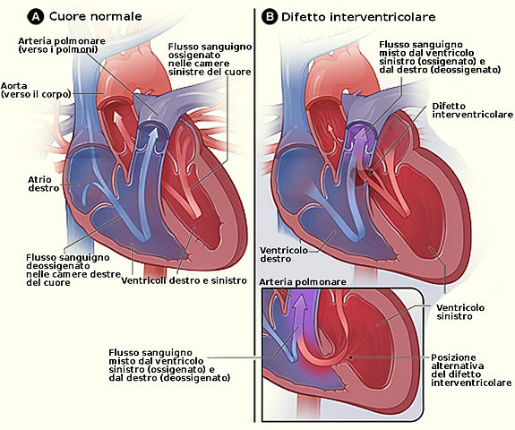
English: Figure A shows the structure and blood flow in the interior of a normal heart. Figure B shows two common locations for a ventricular septal defect. The defect allows oxygen-rich blood from the left ventricle to mix with oxygen-poor blood in the right ventricle. |
| Date | (UTC) |
| Source |
This file was derived from: VSD image.jpg: |
| Author |
|
| This is a retouched picture, which means that it has been digitally altered from its original version. Modifications: tr. The original can be viewed here: VSD image.jpg:
|
Licensing
[edit]
This work is in the public domain in the United States because it is a work prepared by an officer or employee of the United States Government as part of that person’s official duties under the terms of Title 17, Chapter 1, Section 105 of the US Code.
Note: This only applies to original works of the Federal Government and not to the work of any individual U.S. state, territory, commonwealth, county, municipality, or any other subdivision. This template also does not apply to postage stamp designs published by the United States Postal Service since 1978. (See § 313.6(C)(1) of Compendium of U.S. Copyright Office Practices). It also does not apply to certain US coins; see The US Mint Terms of Use.
|
 | |
| This file has been identified as being free of known restrictions under copyright law, including all related and neighboring rights. | ||
Original upload log
[edit]
This image is a derivative work of the following images:
- File:VSD_image.jpg licensed with PD-USGov
- 2010-08-02T09:49:05Z Jmh649 475x421 (108634 Bytes) {{Information |Description={{en|1=Figure A shows the structure and blood flow in the interior of a normal heart. Figure B shows two common locations for a ventricular septal defect. The defect allows oxygen-rich blood from th
Uploaded with derivativeFX
Licensing
[edit]
This file is licensed under the Creative Commons Attribution-Share Alike 2.5 Generic license.
- You are free:
- to share – to copy, distribute and transmit the work
- to remix – to adapt the work
- Under the following conditions:
- attribution – You must give appropriate credit, provide a link to the license, and indicate if changes were made. You may do so in any reasonable manner, but not in any way that suggests the licensor endorses you or your use.
- share alike – If you remix, transform, or build upon the material, you must distribute your contributions under the same or compatible license as the original.
File history
Click on a date/time to view the file as it appeared at that time.
| Date/Time | Thumbnail | Dimensions | User | Comment | |
|---|---|---|---|---|---|
| current | 08:06, 1 April 2012 | | 735 × 614 (176 KB) | Adert (talk | contribs) | Fix refusi nel testo |
| 20:00, 31 March 2012 |  | 735 × 614 (176 KB) | Adert (talk | contribs) | == {{int:filedesc}} == {{Information |Description={{en|1=Figure A shows the structure and blood flow in the interior of a normal heart. Figure B shows two common locations for a ventricular septal defect. The defect allows oxygen-rich blood from the le... |
You cannot overwrite this file.
File usage on Commons
There are no pages that use this file.
File usage on other wikis
The following other wikis use this file:
- Usage on it.wikipedia.org
- Usage on sr.wikipedia.org