File:Trinchesia diljuvia (10.1038-s41598-019-42297-5) Figure 10.png
Jump to navigation
Jump to search
Size of this preview: 630 × 599 pixels. Other resolutions: 252 × 240 pixels | 505 × 480 pixels | 808 × 768 pixels | 1,077 × 1,024 pixels | 1,650 × 1,569 pixels.
Original file (1,650 × 1,569 pixels, file size: 1.14 MB, MIME type: image/png)
Captions
Captions
Add a one-line explanation of what this file represents
Summary
[edit]_Figure_10.png&action=edit§ion=1)
| Description |
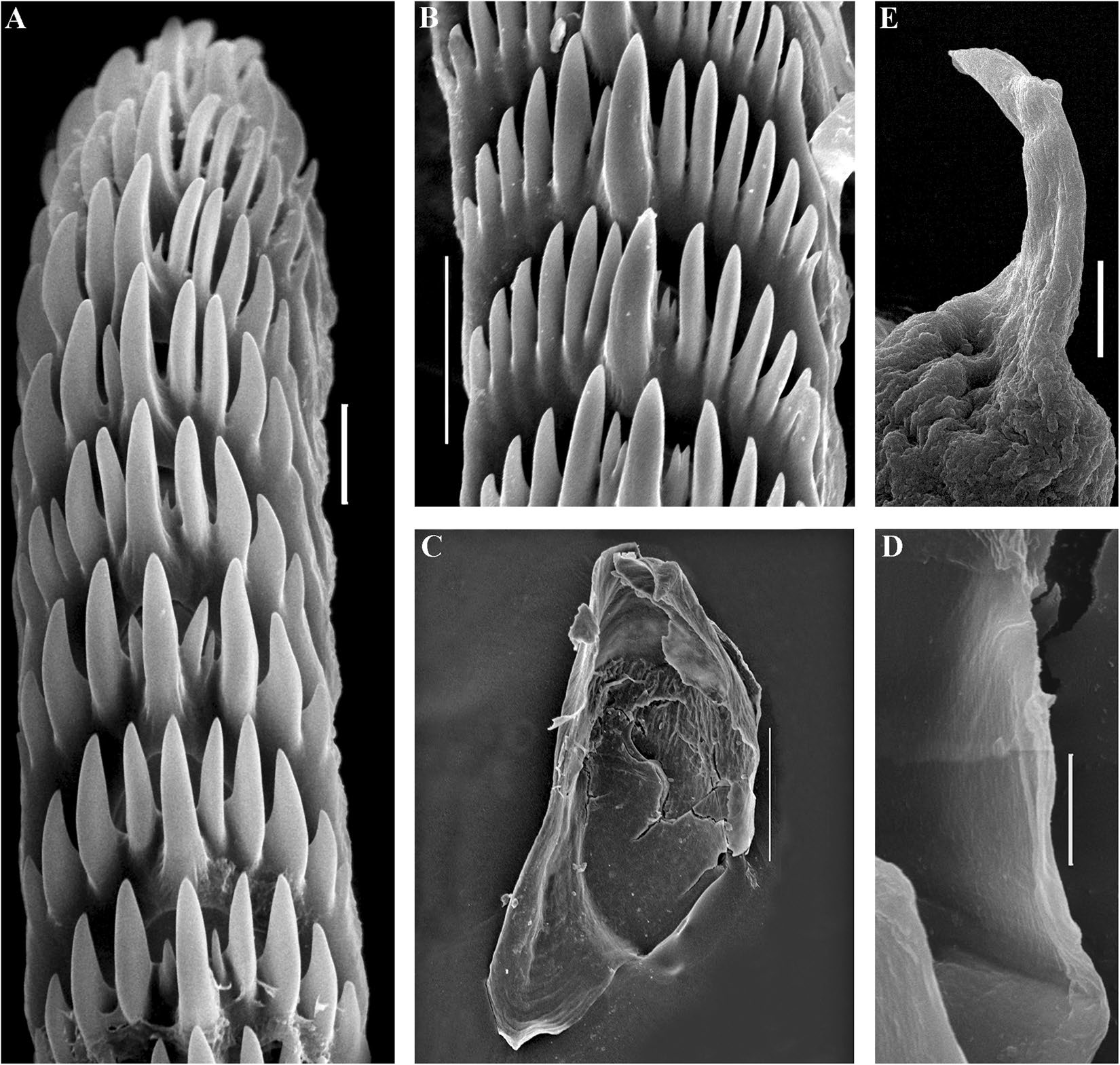
English: Figure 10. Trinchesia diljuvia sp. n. Internal morphology, scanning electron microscopy. (A) Posterior part of radula of paratype ZMMU Op-645; (B). Posterior part of radula (specimen ZMMU Op-643); (C). Jaw, paratype ZMMU Op-643; (D). Details of masticatory processes of jaws, same specimen; (E). Details of copulative organ with stylet, same specimen (ZMMU Op-645). Scale bars: (a), (d), (e) −10 μm; (b) −20 μm; (c) −120 μm. |
| Date | |
| Source | https://dx.doi.org/10.1038%2Fs41598-019-42297-5 |
| Author | [SEM micrographs: Alexander Martynov] Korshunova, T., Picton, B., Furfaro, G., Mariottini, P., Pontes, M., Prkić, J., Fletcher, K., Malmberg, K., Lundin, K., Martynov, A. 2019. Multilevel fine-scale diversity challenges the 'cryptic species' concept. Scientific Reports. 9(6732): 1-21. DOI: 10.1038/s41598-019-42297-5 |
| Permission (Reusing this file) |
This file is licensed under the Creative Commons Attribution 4.0 International license.
|
File history
Click on a date/time to view the file as it appeared at that time.
| Date/Time | Thumbnail | Dimensions | User | Comment | |
|---|---|---|---|---|---|
| current | 18:57, 11 March 2021 | | 1,650 × 1,569 (1.14 MB) | Christian Ferrer (talk | contribs) | {{Information | description = {{en|1=Figure 10. ''Trinchesia diljuvia'' sp. n. Internal morphology, scanning electron microscopy. (A) Posterior part of radula of paratype ZMMU Op-645; (B). Posterior part of radula (specimen ZMMU Op-643); (C). Jaw, paratype ZMMU Op-643; (D). Details of masticatory processes of jaws, same specimen; (E). Details of copulative organ with stylet, same specimen (ZMMU Op-645). Scale bars: (a), (d), (e) −10 μm; (b) −20 μm; (c) −120 μm.}} | date = 2019-05-01 | sour... |
You cannot overwrite this file.
File usage on Commons
There are no pages that use this file.
_Figure_10.png&oldid=659141568)