File:Trinchesia cuanensis (10.1038-s41598-019-42297-5) Figure 6.png
Jump to navigation
Jump to search

Size of this preview: 464 × 599 pixels. Other resolutions: 186 × 240 pixels | 372 × 480 pixels | 595 × 768 pixels | 793 × 1,024 pixels | 1,650 × 2,130 pixels.
Original file (1,650 × 2,130 pixels, file size: 3.11 MB, MIME type: image/png)
Captions
Captions
Add a one-line explanation of what this file represents
Summary
[edit]_Figure_6.png&action=edit§ion=1)
| Description |
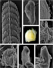
English: Figure 6. Trinchesia cuanensis sp. n. Internal morphology, scanning and light electron microscopy. (A) Posterior part of radula of holotype from Northern Ireland (ZMMU Op-650). (B) Jaw, holotype. (C) Details of masticatory processes of jaws, holotype. (D) Copulative organ with stylet of paratype from Northern Ireland (GNM Gastropoda – 9054), light microscopy. (E) Same, scanning electron microscopy. (F) Details of stylet, same paratype. (G) Details of apical part of stylet, same specimen. (H) Cross section of basal part of stylet, showing channel inside, holotype. Scale bars: a, f −20 μm, b, e −100 μm, c, h, g −10 μm. |
| Date | |
| Source | https://dx.doi.org/10.1038%2Fs41598-019-42297-5 |
| Author | [SEM micrographs: Alexander Martynov] Korshunova, T., Picton, B., Furfaro, G., Mariottini, P., Pontes, M., Prkić, J., Fletcher, K., Malmberg, K., Lundin, K., Martynov, A. 2019. Multilevel fine-scale diversity challenges the 'cryptic species' concept. Scientific Reports. 9(6732): 1-21. DOI: 10.1038/s41598-019-42297-5 |
| Permission (Reusing this file) |
This file is licensed under the Creative Commons Attribution 4.0 International license.
|
File history
Click on a date/time to view the file as it appeared at that time.
| Date/Time | Thumbnail | Dimensions | User | Comment | |
|---|---|---|---|---|---|
| current | 18:51, 11 March 2021 | | 1,650 × 2,130 (3.11 MB) | Christian Ferrer (talk | contribs) | {{Information | description = {{en|1=Figure 6. ''Trinchesia cuanensis'' sp. n. Internal morphology, scanning and light electron microscopy. (A) Posterior part of radula of holotype from Northern Ireland (ZMMU Op-650). (B) Jaw, holotype. (C) Details of masticatory processes of jaws, holotype. (D) Copulative organ with stylet of paratype from Northern Ireland (GNM Gastropoda – 9054), light microscopy. (E) Same, scanning electron microscopy. (F) Details of stylet, same paratype. (G) Details of... |
You cannot overwrite this file.
File usage on Commons
There are no pages that use this file.
_Figure_6.png&oldid=659141898)