File:Trigeminal nerve neurovascular conflict.JPG
Jump to navigation
Jump to search
No higher resolution available.
Trigeminal_nerve_neurovascular_conflict.JPG (768 × 576 pixels, file size: 262 KB, MIME type: image/jpeg)
Captions
Captions
Add a one-line explanation of what this file represents
Summary
[edit]
| Description |
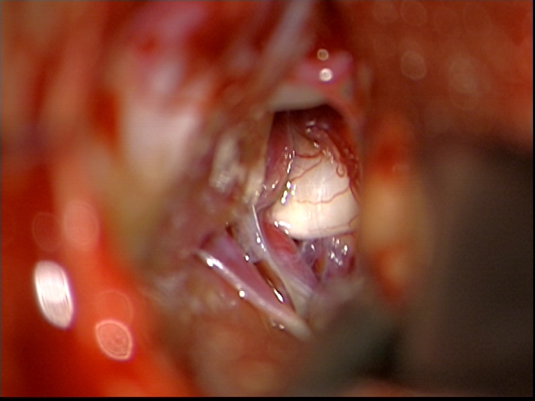
English: Nervo trigemino circondato e compresso da vasi sanguigni (conflitto neuro-vascolare). Foto ottenuta con microscopio operatorio ad elevato ingrandimento durante la fase iniziale di un intervento di microdecompressione vascolare per nevralgia del trigemino.
Trigeminal nerve folded and compressed by blood vessels (neuro-vascular conflict). This photo was obtained with surgical microscope at the early stage of a microvascular decompression operation for trigeminal neuralgia. Trigeminal nerve, visualized with surgical microscope (14 x) during an operation of microvascular decompression (MVD). After accessing the skull base behind the ear and gentle retraction of the cerebellar hemisphere, the trigeminal nerve is visualized at its origin from the brainstem, in the deepest portion of the brain. The compression from vessels - named neuro-vascular conflict - causes trigeminal neuralgia that is an extremely painful and disabling syndrome. The operation consists of resolution of neuro-vascular conflict by mobilization of the offending vessels and leads to resolution of the pain syndrome.English: Trigeminal nerve folded and compressed by blood vessels (neuro-vascular conflict). This photo was obtained with surgical microscope at the early stage of a microvascular decompression operation for trigeminal neuralgia. |
| Date | |
| Source | Own work |
| Author | Luigi Berra |
Licensing
[edit]
I, the copyright holder of this work, hereby publish it under the following license:
This file is licensed under the Creative Commons Attribution-Share Alike 4.0 International license.
- You are free:
- to share – to copy, distribute and transmit the work
- to remix – to adapt the work
- Under the following conditions:
- attribution – You must give appropriate credit, provide a link to the license, and indicate if changes were made. You may do so in any reasonable manner, but not in any way that suggests the licensor endorses you or your use.
- share alike – If you remix, transform, or build upon the material, you must distribute your contributions under the same or compatible license as the original.
| This image was uploaded as part of European Science Photo Competition 2015. |
File history
Click on a date/time to view the file as it appeared at that time.
| Date/Time | Thumbnail | Dimensions | User | Comment | |
|---|---|---|---|---|---|
| current | 18:30, 21 November 2015 | | 768 × 576 (262 KB) | Luigiberra (talk | contribs) | User created page with UploadWizard |
You cannot overwrite this file.
File usage on Commons
There are no pages that use this file.
File usage on other wikis
The following other wikis use this file:
- Usage on it.wikipedia.org
- Usage on no.wikipedia.org
- Usage on www.wikidata.org