File:Transverse sections through the anlage of the right Müllerian duct from a 10 mm. human embryo. X 250.png

Original file (1,009 × 751 pixels, file size: 349 KB, MIME type: image/png)
Captions
Captions
Summary
[edit]
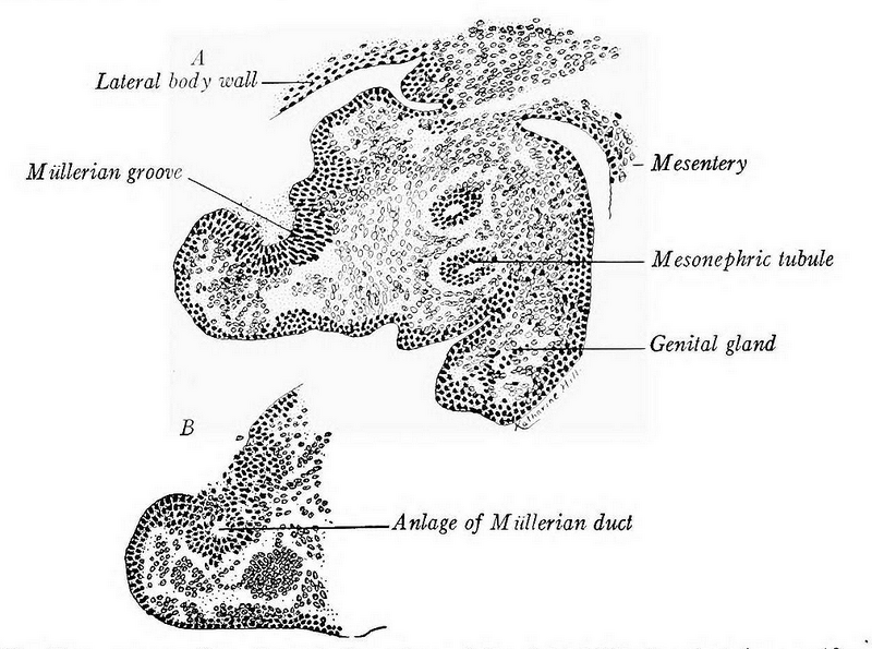
| Description | Fig. 222.—Transverse sections through the anlage of the right Müllerian duct from a 10 mm. human embryo. X 250. A, showing the groove in the urogenital epithelium; B, three sections caudad showing the tubular anlage of the duct. |
| Date | |
| Source | https://www.biodiversitylibrary.org/item/117851#page/227/mode/1up https://www.biodiversitylibrary.org/item/117851#page/7/mode/1up A laboratory manual and text-book of embryology. Philadelphia and London, W. B. Saunders company, 1917. Edition 2d ed., enl. with 388 illustrations, many in color. DOI: https://doi.org/10.5962/bhl.title.56991 |
| Author | Prentiss, Charles William, 1874-1915, Arey, Leslie Brainerd, 1891- |

|
This file, which was originally posted to an external website, has not yet been reviewed by an administrator or reviewer to confirm that the above license is valid. See Category:License review needed for further instructions.
|
Licensing
[edit]
|
This work is in the public domain in its country of origin and other countries and areas where the copyright term is the author's life plus 70 years or fewer.
| |
| This file has been identified as being free of known restrictions under copyright law, including all related and neighboring rights. | |
File history
Click on a date/time to view the file as it appeared at that time.
| Date/Time | Thumbnail | Dimensions | User | Comment | |
|---|---|---|---|---|---|
| current | 20:35, 23 July 2024 | | 1,009 × 751 (349 KB) | Rasbak (talk | contribs) | {{Information |description= Fig. 222.—Transverse sections through the anlage of the right Müllerian duct from a 10 mm. human embryo. X 250. A, showing the groove in the urogenital epithelium; B, three sections caudad showing the tubular anlage of the duct. |date = 1917 |source=https://www.biodiversitylibrary.org/item/117851#page/227/mode/1up https://www.biodiversitylibrary.org/item/117851#page/7/mode/1up A laboratory manual and text-book of embryology. Philadelphia and London, W. B. Saunder... |
You cannot overwrite this file.
File usage on Commons
There are no pages that use this file.