File:Transverse plane of the FT fetal abdomen human.jpg
Jump to navigation
Jump to search

Size of this preview: 800 × 403 pixels. Other resolutions: 320 × 161 pixels | 640 × 323 pixels | 1,024 × 516 pixels | 1,280 × 646 pixels | 2,776 × 1,400 pixels.
Original file (2,776 × 1,400 pixels, file size: 2.15 MB, MIME type: image/jpeg)
Captions
Captions
Add a one-line explanation of what this file represents
Summary
[edit]
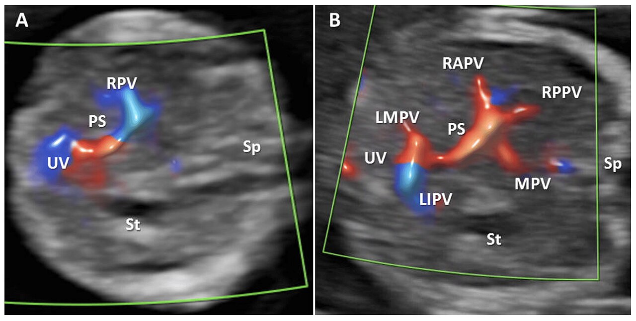
| Description | Figure 2. Transverse plane of the FT fetal abdomen, with high-definition directional power Doppler applied. (A): Transabdominal approach demonstrating the normal L-shaped UV confluence. (B): Transabdominal approach demonstrating all PVS features. MPV main portal vein, PS portal sinus, UV umbilical vein, RAPV anterior branch of right portal vein, RPPV posterior branch of right portal vein, LIPV left portal vein inferior branch; LMPV left portal vein medial branch, St stomach, Ao aorta, Sp spine. |
| Date | |
| Source | Feasibility of Fetal Portal Venous System Ultrasound Assessment at the FT Anomaly Scan. Diagnostics 2022, 12, 361. https://doi.org/10.3390/diagnostics12020361 |
| Author | Nagy, R.D.; Ruican, D.; Zorilă, G.-L.; Istrate-Ofiţeru, A.-M.; Badiu, A.M.; Iliescu, D.G. |
Open acces
Licensing
[edit]
This file is licensed under the Creative Commons Attribution 4.0 International license.
- You are free:
- to share – to copy, distribute and transmit the work
- to remix – to adapt the work
- Under the following conditions:
- attribution – You must give appropriate credit, provide a link to the license, and indicate if changes were made. You may do so in any reasonable manner, but not in any way that suggests the licensor endorses you or your use.

|
This file, which was originally posted to an external website, has not yet been reviewed by an administrator or reviewer to confirm that the above license is valid. See Category:License review needed for further instructions.
|
File history
Click on a date/time to view the file as it appeared at that time.
| Date/Time | Thumbnail | Dimensions | User | Comment | |
|---|---|---|---|---|---|
| current | 15:40, 15 February 2024 | | 2,776 × 1,400 (2.15 MB) | Rasbak (talk | contribs) | {{Information |description=Figure 2. Transverse plane of the FT fetal abdomen, with high-definition directional power Doppler applied. (A): Transabdominal approach demonstrating the normal L-shaped UV confluence. (B): Transabdominal approach demonstrating all PVS features. MPV main portal vein, PS portal sinus, UV umbilical vein, RAPV anterior branch of right portal vein, RPPV posterior branch of right portal vein, LIPV left portal vein inferior branch; LMPV left portal vein medial branch, St... |
You cannot overwrite this file.
File usage on Commons
There are no pages that use this file.