File:Transver cells fpls-05-00046-g006.jpg
Jump to navigation
Jump to search

Size of this preview: 495 × 599 pixels. Other resolutions: 198 × 240 pixels | 396 × 480 pixels | 645 × 781 pixels.
Original file (645 × 781 pixels, file size: 389 KB, MIME type: image/jpeg)
Captions
Captions
Add a one-line explanation of what this file represents
Summary
[edit]
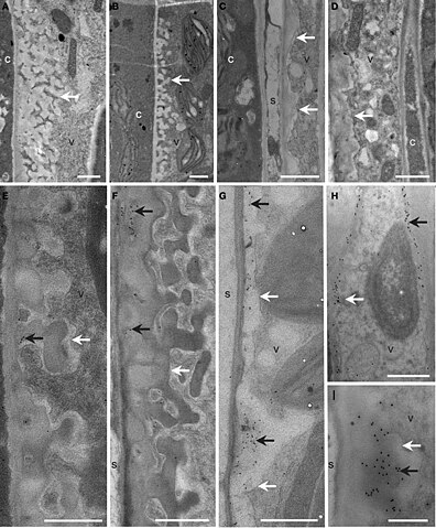
| Description | Figure 6. Cellular structure and immunodetection of callose after 3 days of LT treatment. Col, gsl5, vte2, and gsl5 vte2 were grown under permissive conditions for 4 weeks and transferred to LT conditions for 3 additional days. Col (A,E), gsl5 (B,F), vte2 (C,G), and gsl5 vte2 (D,H,I). Black arrows highlight wall ingrowths of phloem parenchyma transfer cells immunolabeled with anti-β-1,3-glucan. White arrows mark transfer cell walls. c, companion cell; s, sieve element; v, vascular parenchyma transfer cell. Bars = 1 μm (A–H), 0.5 μm (I). |
| Date | |
| Source | https://www.frontiersin.org/files/Articles/76650/fpls-05-00046-HTML/image_m/fpls-05-00046-g006.jpg https://www.frontiersin.org/articles/10.3389/fpls.2014.00046/full Role of callose synthases in transfer cell wall development in tocopherol deficient Arabidopsis mutants, Front. Plant Sci., 19 February 2014, Sec. Plant Physiology, Volume 5 - 2014 |
| Author | Hiroshi Maeda1, Wan Song1, Tammy Sage, Dean DellaPenna1 |
- Error in {{Information}} template: unknown parameter "1".
Open access
Licensing
[edit]
This file is licensed under the Creative Commons Attribution 3.0 Unported license.
- You are free:
- to share – to copy, distribute and transmit the work
- to remix – to adapt the work
- Under the following conditions:
- attribution – You must give appropriate credit, provide a link to the license, and indicate if changes were made. You may do so in any reasonable manner, but not in any way that suggests the licensor endorses you or your use.
File history
Click on a date/time to view the file as it appeared at that time.
| Date/Time | Thumbnail | Dimensions | User | Comment | |
|---|---|---|---|---|---|
| current | 18:23, 1 January 2024 | | 645 × 781 (389 KB) | Rasbak (talk | contribs) | {{Information |description=Figure 6. Cellular structure and immunodetection of callose after 3 days of LT treatment. Col, gsl5, vte2, and gsl5 vte2 were grown under permissive conditions for 4 weeks and transferred to LT conditions for 3 additional days. Col (A,E), gsl5 (B,F), vte2 (C,G), and gsl5 vte2 (D,H,I). Black arrows highlight wall ingrowths of phloem parenchyma transfer cells immunolabeled with anti-β-1,3-glucan. White arrows mark transfer cell walls. c, companion cell; s, sieve eleme... |
You cannot overwrite this file.
File usage on Commons
There are no pages that use this file.
File usage on other wikis
The following other wikis use this file:
- Usage on nl.wikipedia.org