File:Three main variations of the venous anatomy of the dural sinuses.PNG
Jump to navigation
Jump to search

Size of this preview: 800 × 564 pixels. Other resolutions: 320 × 226 pixels | 640 × 451 pixels | 1,024 × 722 pixels | 1,280 × 902 pixels | 1,521 × 1,072 pixels.
Original file (1,521 × 1,072 pixels, file size: 385 KB, MIME type: image/png)
Captions
Captions
Add a one-line explanation of what this file represents
Summary
[edit]
| Description |
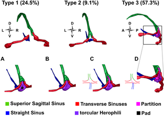
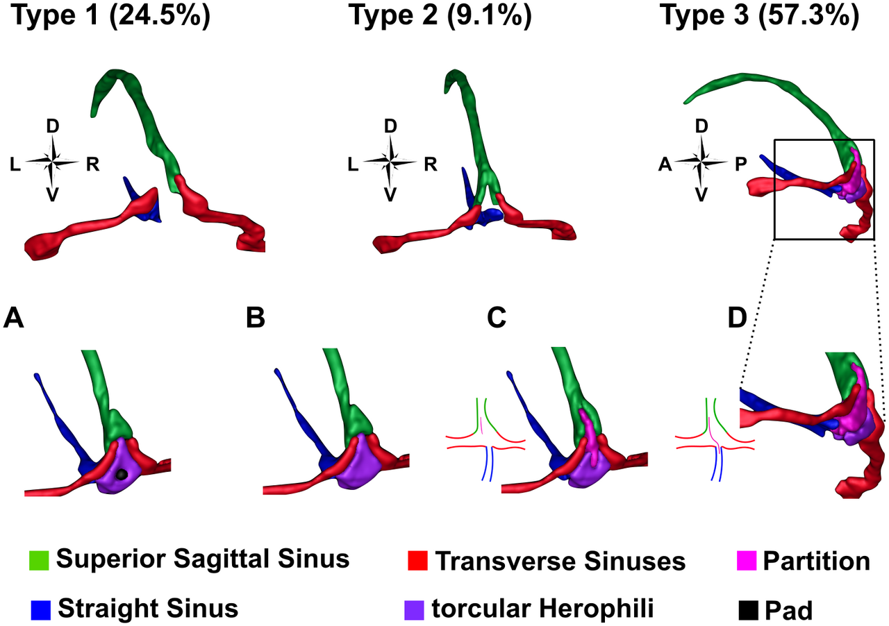
English: Three main variations of the venous anatomy of the dural sinuses.
Percentages refer to the frequency with which each type is seen. In Type 1, the Superior Sagittal Sinus connects to one Transverse Sinus and the Straight Sinus to the other, with the two TSs completely separate from one another. In Type 2, the SSS and SS are forked, with the left forks connecting to the left TS, and the right forks to the right TS. In Type 3 there is some variation of a confluence of the sinuses. A) A ‘pad-like dural elevation’ in the tH [54]. B) Sinuses are connected by a confluence at the occipital pole. C) A partial partition extends from the SSS into the confluence of sinuses. This partition can be closer to the left or the right wall of the SSS. D) A full partition extends from the SSS diagonally through the tH to the SS. The full partition can extend diagonally across from closer to the left or the right wall of the SSS. This figure was created by segmenting the venous anatomy from the venogram of Subject 4, who had Type 3A. Other variations were approximated based on the descriptions in [54]. |
| Date | |
| Source | Boyd Taylor HG, Puckett AM, Isherwood ZJ, Schira MM (2019) Vascular effects on the BOLD response and the retinotopic mapping of hV4. PLoS ONE 14(6): e0204388. https://doi.org/10.1371/journal.pone.0204388 |
| Author | H. G. Boyd Taylor , A. M. Puckett, Z. J. Isherwood, M. M. Schira |
Licensing
[edit]
This file is licensed under the Creative Commons Attribution 4.0 International license.
- You are free:
- to share – to copy, distribute and transmit the work
- to remix – to adapt the work
- Under the following conditions:
- attribution – You must give appropriate credit, provide a link to the license, and indicate if changes were made. You may do so in any reasonable manner, but not in any way that suggests the licensor endorses you or your use.
File history
Click on a date/time to view the file as it appeared at that time.
| Date/Time | Thumbnail | Dimensions | User | Comment | |
|---|---|---|---|---|---|
| current | 17:35, 6 July 2019 | | 1,521 × 1,072 (385 KB) | Was a bee (talk | contribs) | {{Information |Description={{en|1=Three main variations of the venous anatomy of the dural sinuses. Percentages refer to the frequency with which each type is seen. In Type 1, the Superior Sagittal Sinus connects to one Transverse Sinus and the Straight Sinus to the other, with the two TSs completely separate from one another. In Type 2, the SSS and SS are forked, with the left forks connecting to the left TS, and the right forks to the right TS. In Type 3 there is some variation of a conflu... |
You cannot overwrite this file.
File usage on Commons
There are no pages that use this file.