File:The chemical neuroanatomy of the brainstem reticular formation (BRF).png
Jump to navigation
Jump to search

Size of this preview: 800 × 395 pixels. Other resolutions: 320 × 158 pixels | 640 × 316 pixels | 1,024 × 505 pixels | 1,958 × 966 pixels.
Original file (1,958 × 966 pixels, file size: 806 KB, MIME type: image/png)
Captions
Captions
Add a one-line explanation of what this file represents
Summary
[edit].png&action=edit§ion=1)
| Description |
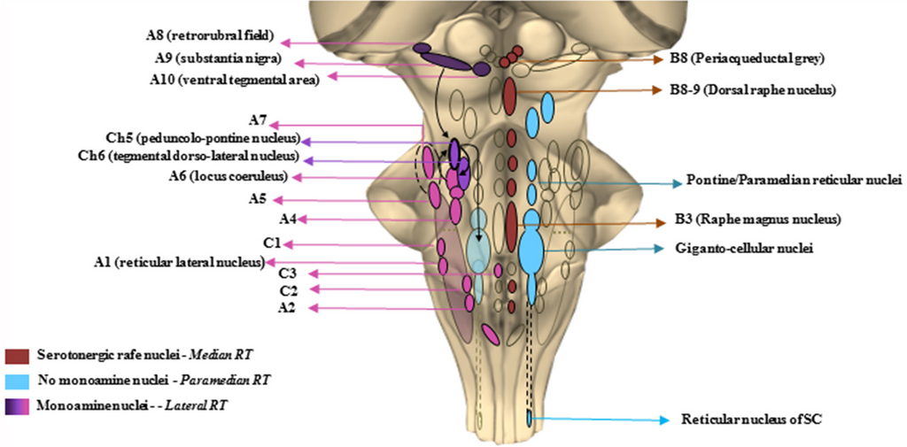
English: The chemical neuroanatomy of the brainstem reticular formation (BRF). This cartoon offers a schematic description of those brainstem areas properly belonging to the reticular formation (RF). In the The diagram shows the constellation of the RF nuclei following a neurotransmitter chemical classification. The isodendritic morphology of the neurons composing the RF nuclei, configures them as crucial stations of both afferent and efferent projections descending and projecting up to the cortex and spinal cord (SC). This network of overlapping connections is involved in a plenty of either extrapyramidal motor and non-motor functions. The major monoamine containing areas, mainly localized in the lateral RF except from C3, are the noradrenergic (A1–A7) adrenergic (C1–C3) dopaminergic (A8–A10) and cholinergic (Ch5–Ch6) nuclei. These are crucial for respiratory activity and for regulating blood pressure and heart rate, micturition, sweat, sleep-wakink cycle as well as descending motor control. Serotonergic nuclei are found in the median RF raphe nuclei, mainly in the B3, B8 and B9 areas. They control vegetative functions such as mood, sleep and sexual behavior, depression and pain. The medial RF, found between the median and the lateral column, is a region lacking monoamine nuclei, but whose giganto-cellular and paramedianpontine nuclei act as a station for fibers connecting with monoamine regions such as A6 (LC) and Ch6. They are involved in voluntary movement regulation, as well as in optical, acoustic and olfactory control due to their connections respectively, to the spinal cord and to the main cranial nerves’ nuclei. |
| Date | Published: 18 April 2017. |
| Source | Gambardella, S., Ferese, R., Biagioni, F., Busceti, C. L., Campopiano, R., Griguoli, A., Limanaqi, F., Novelli, G., Storto, M., & Fornai, F. (2017). The Monoamine Brainstem Reticular Formation as a Paradigm for Re-Defining Various Phenotypes of Parkinson's Disease Owing Genetic and Anatomical Specificity. Frontiers in cellular neuroscience, 11, 102. https://doi.org/10.3389/fncel.2017.00102 |
| Author | Stefano Gambardella, Rosangela Ferese, Francesca Biagioni, Carla L. Busceti, Rosa Campopiano, Anna M. P. Griguoli, Fiona Limanaqi, Giuseppe Novelli, Marianna Storto, and Francesco Fornai |
Licensing
[edit].png&action=edit§ion=2)
This file is licensed under the Creative Commons Attribution 4.0 International license.
- You are free:
- to share – to copy, distribute and transmit the work
- to remix – to adapt the work
- Under the following conditions:
- attribution – You must give appropriate credit, provide a link to the license, and indicate if changes were made. You may do so in any reasonable manner, but not in any way that suggests the licensor endorses you or your use.
File history
Click on a date/time to view the file as it appeared at that time.
| Date/Time | Thumbnail | Dimensions | User | Comment | |
|---|---|---|---|---|---|
| current | 12:55, 1 December 2020 | | 1,958 × 966 (806 KB) | Was a bee (talk | contribs) | {{Information |Description={{en|1=The chemical neuroanatomy of the brainstem reticular formation (BRF). This cartoon offers a schematic description of those brainstem areas properly belonging to the reticular formation (RF). In the The diagram shows the constellation of the RF nuclei following a neurotransmitter chemical classification. The isodendritic morphology of the neurons composing the RF nuclei, configures them as crucial stations of both afferent and efferent projections descending a... |
You cannot overwrite this file.
File usage on Commons
There are no pages that use this file.
File usage on other wikis
The following other wikis use this file:
- Usage on en.wikiversity.org
- User:Jtwsaddress42/Projects/Project 5
- User:Jtwsaddress42/Projects/Project 5/Parts
- User:Jtwsaddress42/Projects/Project 5/Parts/Part 1
- User:Jtwsaddress42/Projects/Project 5/Chapters/Chapter 1
- User:Jtwsaddress42/Projects/Project 5/Sections/Chapter 1/Alfred Sherwood Romer - The Vertebrate as a Dual Organism
.png&oldid=624059708)