File:TAAR1 Amphetamine Dopamine.png
Jump to navigation
Jump to search
Size of this preview: 472 × 599 pixels. Other resolutions: 189 × 240 pixels | 378 × 480 pixels | 807 × 1,024 pixels.
Original file (807 × 1,024 pixels, file size: 188 KB, MIME type: image/png)
Captions
Captions
Add a one-line explanation of what this file represents
Summary
[edit]| Description |
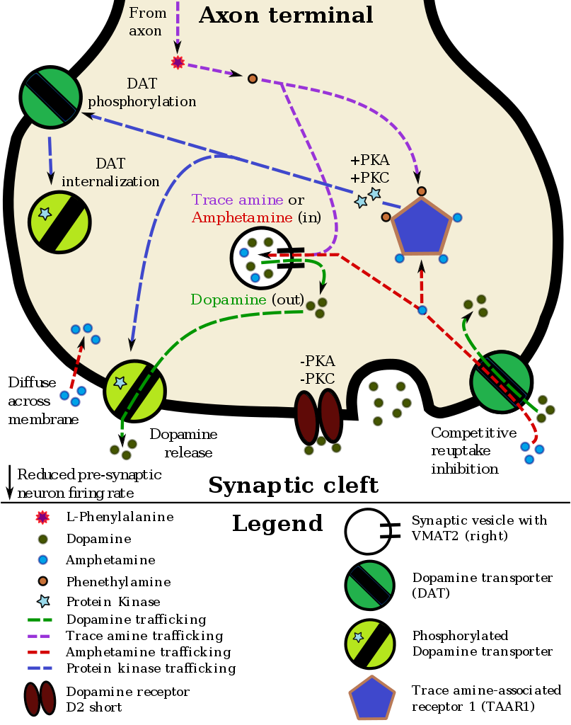
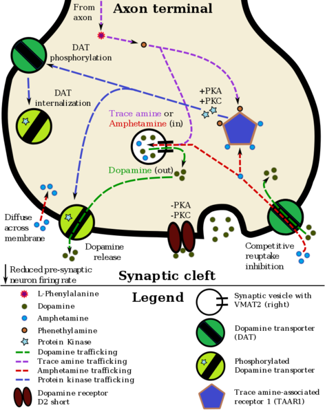
English: This is an illustration of a dopamine neuron with co-localized TAAR1 and the effects of a TAAR1 agonist (amphetamine or a trace amine) on dopamine reuptake and effluxion.
This model was based upon information from the three following sources:
|
|||
| Date | ||||
| Source | Own work | |||
| Author | Seppi333 | |||
| Other versions |
|


Licensing
[edit]
I, the copyright holder of this work, hereby publish it under the following license:
This file is licensed under the Creative Commons Attribution-Share Alike 3.0 Unported license.
- You are free:
- to share – to copy, distribute and transmit the work
- to remix – to adapt the work
- Under the following conditions:
- attribution – You must give appropriate credit, provide a link to the license, and indicate if changes were made. You may do so in any reasonable manner, but not in any way that suggests the licensor endorses you or your use.
- share alike – If you remix, transform, or build upon the material, you must distribute your contributions under the same or compatible license as the original.
File history
Click on a date/time to view the file as it appeared at that time.
| Date/Time | Thumbnail | Dimensions | User | Comment | |
|---|---|---|---|---|---|
| current | 04:23, 4 September 2017 | | 807 × 1,024 (188 KB) | Seppi333 (talk | contribs) | Uploading small fix to a line of text in this image (this is the update from the April 2017 revision of File:TAAR1_Dopamine.svg) |
| 07:05, 29 January 2016 |  | 807 × 1,024 (202 KB) | Seppi333 (talk | contribs) | fix from svg version | |
| 08:35, 17 April 2015 |  | 807 × 1,024 (202 KB) | Seppi333 (talk | contribs) | new version w/ different cytosol color | |
| 00:35, 22 May 2014 |  | 725 × 920 (179 KB) | Seppi333 (talk | contribs) | rv to SVG variant | |
| 19:50, 23 March 2014 |  | 4,028 × 5,111 (1.39 MB) | Seppi333 (talk | contribs) | increase size, update content for review by others before updating svg file | |
| 20:11, 15 March 2014 |  | 725 × 920 (179 KB) | Seppi333 (talk | contribs) | update to current svg version | |
| 14:09, 25 February 2014 |  | 740 × 925 (173 KB) | Seppi333 (talk | contribs) | Uploading png version of the new svg file | |
| 05:28, 11 January 2014 |  | 800 × 975 (85 KB) | Seppi333 (talk | contribs) | Added dopamine to synaptic cleft side of the D2 receptor. Refined minor details (e.g., certain arrowheads). | |
| 02:35, 15 December 2013 |  | 800 × 975 (85 KB) | Seppi333 (talk | contribs) | Added post-synaptic DA firing-rate reduction - while not indicated in the picture, this is TAAR1-mediated, presumably through an effect on adjacent inhibitory neurons. (See Miller, FIG 1) Also move "pre-synaptic" and "synaptic cleft" more to the right. | |
| 02:43, 13 December 2013 |  | 800 × 975 (82 KB) | Seppi333 (talk | contribs) | Extremely minute detail fix of the gray arrow through DAT. |
You cannot overwrite this file.
File usage on Commons
There are no pages that use this file.
File usage on other wikis
The following other wikis use this file:
- Usage on en.wikipedia.org
- Usage on fr.wikipedia.org