File:Syringomyelia2.jpg
Jump to navigation
Jump to search
No higher resolution available.
Syringomyelia2.jpg (612 × 508 pixels, file size: 36 KB, MIME type: image/jpeg)
Captions
Captions
Add a one-line explanation of what this file represents
Summary
[edit]
| Description |
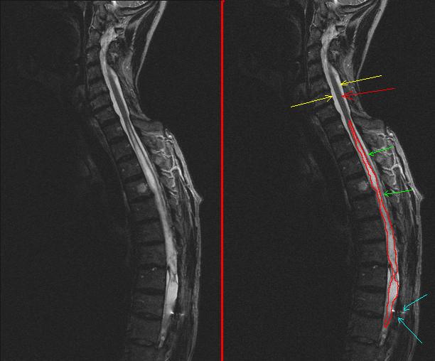
English: MRI image of the sagital thoracic spine with syringomyelia
Nederlands: MRI-beeld van een sagittale doorsnede van de rug ter hoogte van de borstwervels met een syringomyelie. (Zie rode omlijning op het rechter plaatje.) De voorkant van de patiënt is hier links op de afbeelding. Boven de afwijking is het ruggenmerg normaal (rode pijl). Rondom het ruggenmerg is er daar voldoende ruimte, hierin bevindt zich het hersenvocht, op deze afbeelding wit. (Gele pijlen). Ter hoogte van de afwijking is er nog maar een dun schilletje van het ruggenmerg over, omdat het centrale kanaal hier erg wijd en gevuld is met hersenvocht (groene pijlen). Overigens is deze patiënt al een keer geopereerd, o.a. te zien aan een metaalartefact (blauwe pijlen). |
| Date | 3 August 2008 (upload date) |
| Source | Own work |
| Author | Lucien Monfils |
On the left side the original image, on the right side the same image with annotations. For explanation of the annotations see the Dutch Wikipedia, or contact me by email.
Links het originele beeld, rechts hetzelfde beeld met annotaties.
Licensing
[edit]
I, the copyright holder of this work, hereby publish it under the following licenses:

|
Permission is granted to copy, distribute and/or modify this document under the terms of the GNU Free Documentation License, Version 1.2 or any later version published by the Free Software Foundation; with no Invariant Sections, no Front-Cover Texts, and no Back-Cover Texts. A copy of the license is included in the section entitled GNU Free Documentation License. |
This file is licensed under the Creative Commons Attribution-Share Alike 3.0 Unported, 2.5 Generic, 2.0 Generic and 1.0 Generic license.
- You are free:
- to share – to copy, distribute and transmit the work
- to remix – to adapt the work
- Under the following conditions:
- attribution – You must give appropriate credit, provide a link to the license, and indicate if changes were made. You may do so in any reasonable manner, but not in any way that suggests the licensor endorses you or your use.
- share alike – If you remix, transform, or build upon the material, you must distribute your contributions under the same or compatible license as the original.
You may select the license of your choice.
File history
Click on a date/time to view the file as it appeared at that time.
| Date/Time | Thumbnail | Dimensions | User | Comment | |
|---|---|---|---|---|---|
| current | 13:50, 3 August 2008 | | 612 × 508 (36 KB) | Lucien Monfils (talk | contribs) | {{Information |Description={{en|1=MRI image of the sagital thoracic spine with syringomyelia}} {{nl|1=MRI beeld van de sagitale thoracele wervelkolom met syringomyelie}} |Source=eigen werk (own work) |Author=Lucien Monfils |Date=03 |
You cannot overwrite this file.
File usage on Commons
The following page uses this file:
File usage on other wikis
The following other wikis use this file:
- Usage on en.wikipedia.org
- Usage on nl.wikipedia.org