File:Sus domesticus blastocyst.jpg
Original file (1,016 × 759 pixels, file size: 647 KB, MIME type: image/jpeg)
Captions
Captions
Summary
[edit]
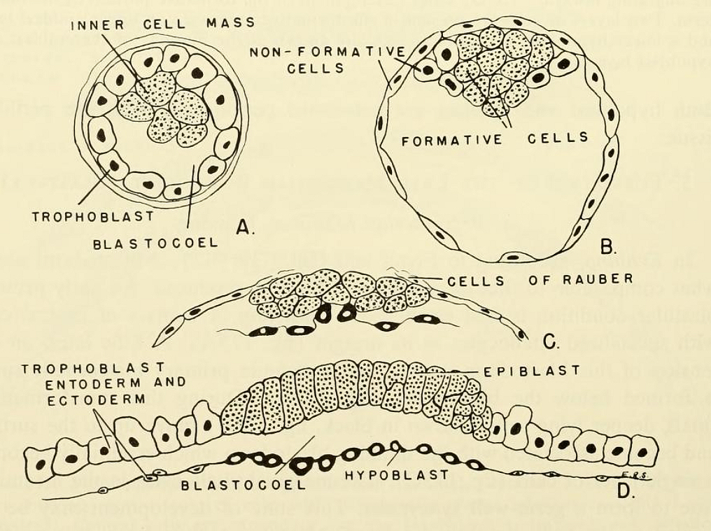
| Description | Fig. 177. Schematic drawings of early pig development. (A) Early developing blastocyst. (B) Later blastocyst, showing two kinds of cells in the inner cell mass. (C) Later blastocyst, showing disappearance of trophoblast cells overlying the inner cell mass. (D) Later blastocyst. Two layers of formative cells are present as indicated with trophoblast tissue attached at the margins. |
| Date | |
| Source | https://archive.org/details/comparativeembry00nels/page/364/mode/1up?view=theater&q=BLASTULATION+ Comparative embryology of the vertebrates; with 2057 drawings and photos. grouped as 380 illustrations. |
| Author | Nelsen, Olin E. |
Licensing
[edit]
|
This work is in the public domain in its country of origin and other countries and areas where the copyright term is the author's life plus 70 years or fewer.
| |
| This file has been identified as being free of known restrictions under copyright law, including all related and neighboring rights. | |

|
This file, which was originally posted to an external website, has not yet been reviewed by an administrator or reviewer to confirm that the above license is valid. See Category:License review needed for further instructions.
|
File history
Click on a date/time to view the file as it appeared at that time.
| Date/Time | Thumbnail | Dimensions | User | Comment | |
|---|---|---|---|---|---|
| current | 10:59, 12 March 2024 | | 1,016 × 759 (647 KB) | Rasbak (talk | contribs) | {{Information |description=Fig. 177. Schematic drawings of early pig development. (A) Early developing blastocyst. (B) Later blastocyst, showing two kinds of cells in the inner cell mass. (C) Later blastocyst, showing disappearance of trophoblast cells overlying the inner cell mass. (D) Later blastocyst. Two layers of formative cells are present as indicated with trophoblast tissue attached at the margins. |date=1953 |source=https://archive.org/details/comparativeembry00nels/page/361/mode/1up... |
You cannot overwrite this file.
File usage on Commons
There are no pages that use this file.