File:Streblospio eridani (10.11646-zootaxa.4742.1.10) figure 7.jpg
Jump to navigation
Jump to search
Size of this preview: 739 × 600 pixels. Other resolutions: 296 × 240 pixels | 592 × 480 pixels | 946 × 768 pixels | 1,262 × 1,024 pixels | 1,417 × 1,150 pixels.
Original file (1,417 × 1,150 pixels, file size: 769 KB, MIME type: image/jpeg)
Captions
Captions
Add a one-line explanation of what this file represents
Summary
[edit]_figure_7.jpg&action=edit§ion=1)
| Description |
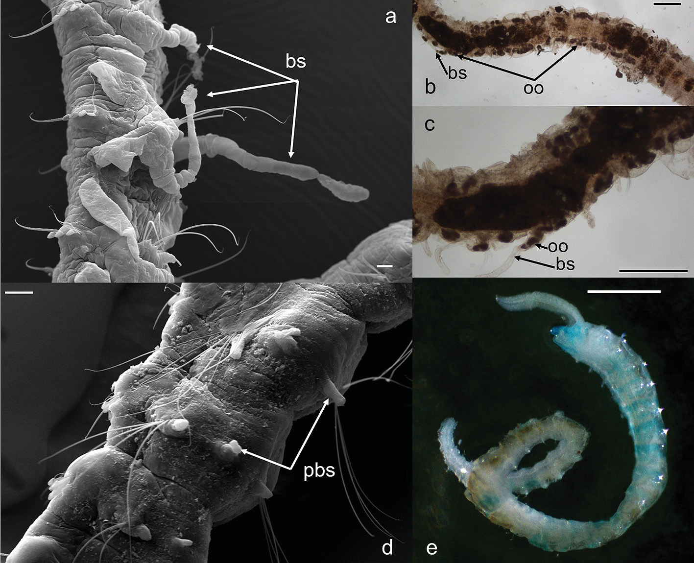
English: FIGURE 7. Streblospio eridani n. sp. a brooding structures. b oocytes along the body, and in a lateral row at each side of the body on anterior chaetigers. c oocytes inside the brooding structures. d posterior branched structures. e methyl green staining pattern (paratype MNHF I.AL.19.0002-9), ventrolateral view Abbreviations: bs, brooding structures; oo, oocytes; pbs, posterior branched structures. Scale bars: a = 20 μm; b-c = 100 μm; d = 30 μm; e = 0.5 mm. |
| Date | |
| Source | https://dx.doi.org/10.11646/zootaxa.4742.1.10 |
| Author | Capa, M. & Bakken, T. 2015. Revision of the Australian Sphaerodoridae (Annelida) including the description of four new species. Zootaxa 4000(2): 227–367. |
| Permission (Reusing this file) |
This file is licensed under the Creative Commons Attribution 4.0 International license.
|
File history
Click on a date/time to view the file as it appeared at that time.
| Date/Time | Thumbnail | Dimensions | User | Comment | |
|---|---|---|---|---|---|
| current | 16:20, 20 July 2021 | | 1,417 × 1,150 (769 KB) | Christian Ferrer (talk | contribs) | {{Information |description={{en|1=FIGURE 7. ''Streblospio eridani'' n. sp. a brooding structures. b oocytes along the body, and in a lateral row at each side of the body on anterior chaetigers. c oocytes inside the brooding structures. d posterior branched structures. e methyl green staining pattern (paratype MNHF I.AL.19.0002-9), ventrolateral view Abbreviations: bs, brooding structures; oo, oocytes; pbs, posterior branched structures. Scale bars: a = 20 μm; b-c = 100 μm; d = 30 μm; e = 0.5... |
You cannot overwrite this file.
File usage on Commons
There are no pages that use this file.
_figure_7.jpg&oldid=576110437)