File:Spinal canal.webp
Jump to navigation
Jump to search
No higher resolution available.
Spinal_canal.webp (707 × 426 pixels, file size: 68 KB, MIME type: image/webp)
Captions
Captions
Add a one-line explanation of what this file represents
Summary
[edit]
| Description |
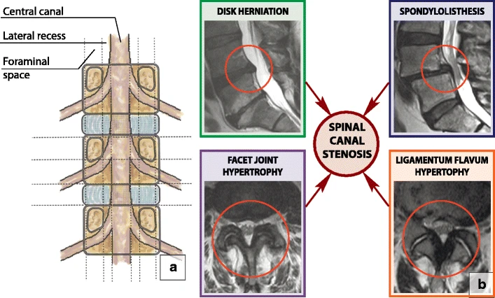
English: Spinal canal. (a) Normal spinal canal. The central portion of the spinal canal is bordered laterally by a lateral recess, dorsally by a vertebral arch and ventrally by a vertebral body and discs. The lateral recess is bordered laterally by a pedicle, dorsally by a superior articular facet and ventrally by a vertebral body and discs. The foraminal space is bordered by cephalad and caudal pedicles and facet joints dorsally and a vertebral body and discs ventrally. The extraforaminal space is lateral to the neuroforamen. (b) Spinal canal stenosis. There are four major causes of degenerative spinal canal stenosis: disc herniation, hypertrophic facet joint osteoarthrosis, ligamentum flavum hypertrophy and degenerative spondylolisthesis |
| Date | |
| Source | Kushchayev, S.V., Glushko, T., Jarraya, M. et al. ABCs of the degenerative spine. Insights Imaging 9, 253–274 (2018). https://doi.org/10.1007/s13244-017-0584-z |
| Author | Irina Nefedova |
Licensing
[edit]
This file is licensed under the Creative Commons Attribution 4.0 International license.
- You are free:
- to share – to copy, distribute and transmit the work
- to remix – to adapt the work
- Under the following conditions:
- attribution – You must give appropriate credit, provide a link to the license, and indicate if changes were made. You may do so in any reasonable manner, but not in any way that suggests the licensor endorses you or your use.
File history
Click on a date/time to view the file as it appeared at that time.
| Date/Time | Thumbnail | Dimensions | User | Comment | |
|---|---|---|---|---|---|
| current | 08:07, 11 April 2021 | | 707 × 426 (68 KB) | Balkanique (talk | contribs) | Uploaded a work by Irina Nefedova from Kushchayev, S.V., Glushko, T., Jarraya, M. et al. ABCs of the degenerative spine. Insights Imaging 9, 253–274 (2018). https://doi.org/10.1007/s13244-017-0584-z with UploadWizard |
You cannot overwrite this file.
File usage on Commons
The following page uses this file: