File:Sphaerodoropsis fauchaldi (10.11646-zootaxa.4000.2.3) Figure 9.jpg
Jump to navigation
Jump to search
Size of this preview: 448 × 599 pixels. Other resolutions: 179 × 240 pixels | 359 × 480 pixels | 574 × 768 pixels | 766 × 1,024 pixels | 1,774 × 2,372 pixels.
Original file (1,774 × 2,372 pixels, file size: 2.24 MB, MIME type: image/jpeg)
Captions
Captions
Add a one-line explanation of what this file represents
Summary
[edit]| Description |
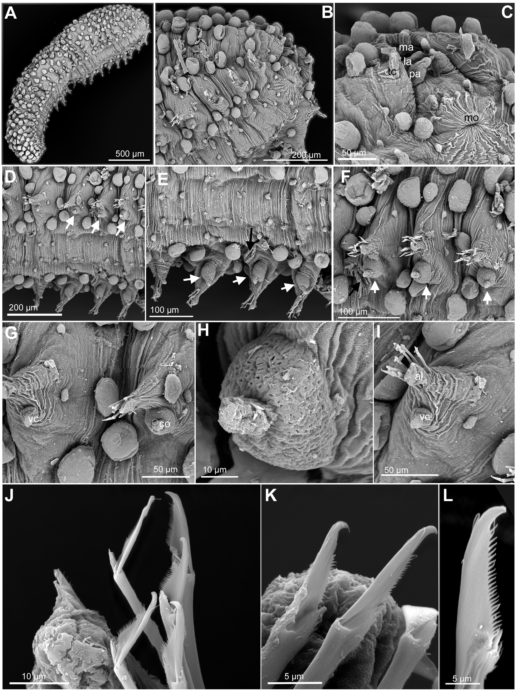
English: FIGURE 9. Sphaerodoropsis fauchaldi, NTM W.10206, scanning electron micrographs. A. Complete specimen, ventro-lateral view. B, Anterior end, ventral view. C. Detail anterior end and head appendages, ventral view. D. Chaetigers 9–5 (left to right), ventral side, with female inflated and porous ventral cirri (white arrows) in chaetigers 7–5. E. Same, opposite side, with inflated and porous ventral cirri (white arrows) in chaetigers 7–5 and oval tubercle (black arrow) on chaetiger 6. F. Parapodia, chaetigers 6–4, with ‘copulatory organs’(arrows). G. Parapodia of chaetigers 8 (with normal ventral cirrus) and 7 (with copulatory organ), antero-lateral view. H. Detail of copulatory organ in chaetiger 6 with pores scattered on the surface. I. Parapodium chaetiger 8, anterior view. J. Chaetal fascicle, mid-body chaetiger. K. Chaetae chaetiger 5. L. Chaeta posterior chaetiger. Abbreviations: al, acicular lobe; co, copulatory organ; la, lateral antenna; ma, median antenna; pa, palp; tc, tentacular cirrus; vc, ventral cirrus. |
| Date | |
| Source | https://dx.doi.org/10.11646%2Fzootaxa.4000.2.3 |
| Author | Capa, M. & Bakken, T. 2015. Revision of the Australian Sphaerodoridae (Annelida) including the description of four new species. Zootaxa 4000(2): 227–367. |
| Permission (Reusing this file) |
This file is licensed under the Creative Commons Attribution 3.0 Unported license.
|
| Other versions |
_Figure_9_(cropped).jpg)
_Figure_9.jpg&action=edit§ion=1)
File history
Click on a date/time to view the file as it appeared at that time.
| Date/Time | Thumbnail | Dimensions | User | Comment | |
|---|---|---|---|---|---|
| current | 16:30, 18 July 2021 | | 1,774 × 2,372 (2.24 MB) | Christian Ferrer (talk | contribs) | {{Information |description={{en|1=FIGURE 9. ''Sphaerodoropsis fauchaldi'', NTM W.10206, scanning electron micrographs. A. Complete specimen, ventro-lateral view. B, Anterior end, ventral view. C. Detail anterior end and head appendages, ventral view. D. Chaetigers 9–5 (left to right), ventral side, with female inflated and porous ventral cirri (white arrows) in chaetigers 7–5. E. Same, opposite side, with inflated and porous ventral cirri (white arrows) in chaetigers 7–5 and oval tubercle (bl... |
You cannot overwrite this file.
File usage on Commons
The following page uses this file:
_Figure_9.jpg&oldid=575779748)