File:Sabellidae (10.11646-zootaxa.4019.1.8) Figure 22.jpg
Jump to navigation
Jump to search
Size of this preview: 427 × 599 pixels. Other resolutions: 171 × 240 pixels | 342 × 480 pixels | 547 × 768 pixels | 730 × 1,024 pixels | 1,688 × 2,368 pixels.
Original file (1,688 × 2,368 pixels, file size: 1.96 MB, MIME type: image/jpeg)
Captions
Captions
Add a one-line explanation of what this file represents
Summary
[edit]| Description |
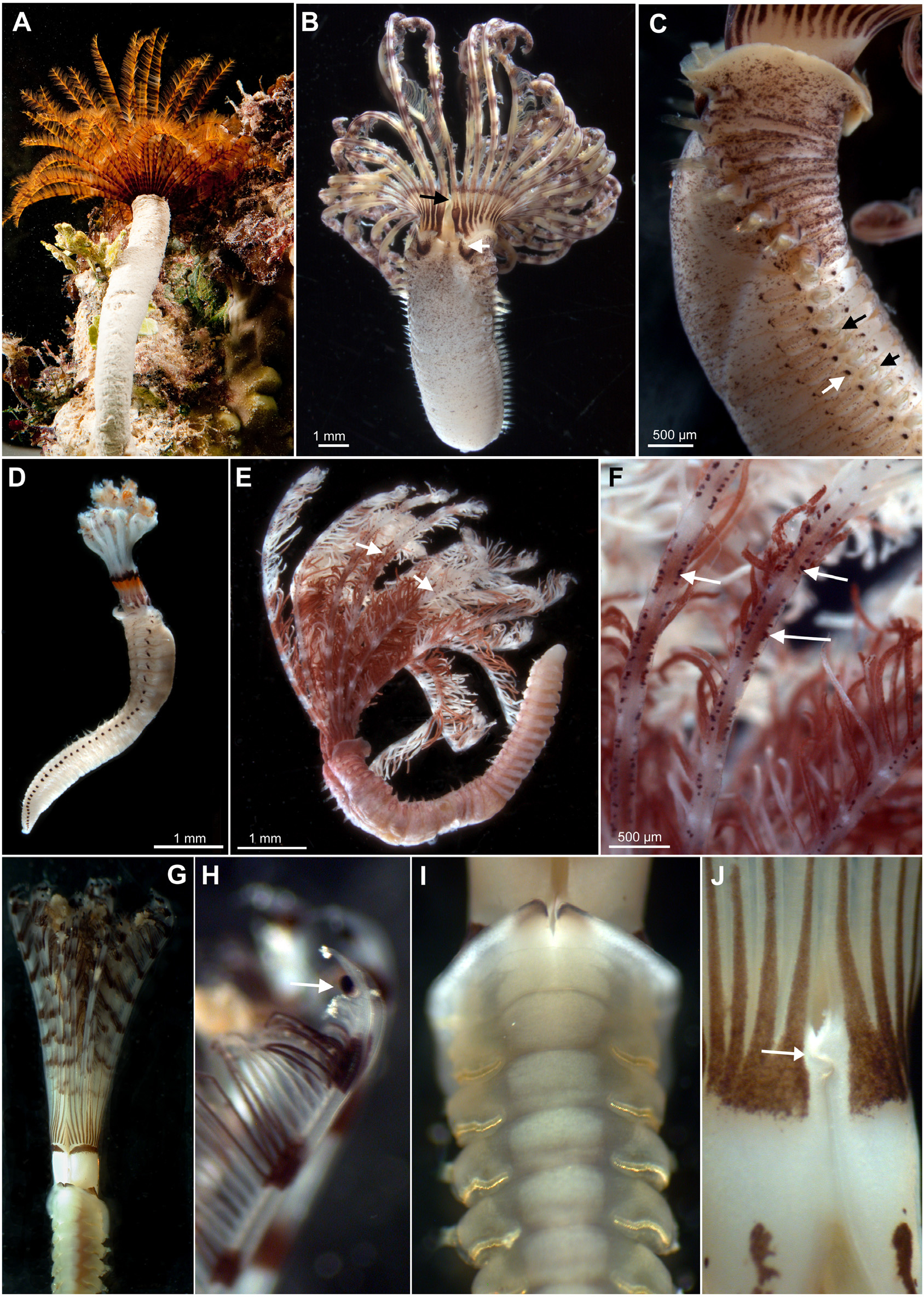
English: FIGURE 22. A–D. Sabellastarte sp. A. Live specimens with radiolar crown exposed outside the tube; B. Dorsal view, showing collar dorsal margins fused to faecal groove forming pockets at either side (white arrow); radiolar lobes with dorsal basal flanges (black arrow); C. Same, lateral view, showing the pigmentation pattern, including the interramal eyespots (white arrow) and the arrangement in a C shape row of inferior abdominal chaetae (black arrows); D. Juvenile showing a different pigmentation pattern to adults; E–F. Sabellomma cupoculata. A. Incomplete specimen, showing pigmentation pattern and rows of ocelli along radiolar margins (arrows); F. Detail of radioles and ocelli (arrows); G–J. Stylomma palmatum, live specimen; G. Anterior end showing the rigid radiolar crown with elongated basal lobes; H. Detail of radiolar tip with unpaired subdistal stalked compound eyes (arrow); I. Anterior abdominal chaetigers, with anterior collar margin incised midventrally and thoracic tori almost in contact with ventral shields; J. Detail of basal lobes, dorsally, showing the flanges joined dorsally with a press stud structure (arrow). |
| Date | |
| Source | https://doi.org/10.11646/zootaxa.4019.1.8 |
| Author | Capa, M. & Murray, A. 2015. A taxonomic guide to the fanworms (Sabellidae, Annelida) of Lizard Island, Great Barrier Reef, Australia, including new species and new records. IN Hutchings, P.A. & Kupriyanova, E.K. (eds.), 2015: Coral reef-associated fauna of Lizard Island, Great Barrier Reef: polychaetes and allies. Zootaxa 4019(1): 98–167. |
| Permission (Reusing this file) |
This file is licensed under the Creative Commons Attribution 3.0 Unported license.
|
| Other versions |
_Figure_22_(cropped).jpg)
_Figure_22_(cropped).jpg)
_Figure_22.jpg&action=edit§ion=1)
File history
Click on a date/time to view the file as it appeared at that time.
| Date/Time | Thumbnail | Dimensions | User | Comment | |
|---|---|---|---|---|---|
| current | 18:10, 22 July 2021 | | 1,688 × 2,368 (1.96 MB) | Christian Ferrer (talk | contribs) | {{Information |description={{en|1=FIGURE 22. A–D. ''Sabellastarte'' sp. A. Live specimens with radiolar crown exposed outside the tube; B. Dorsal view, showing collar dorsal margins fused to faecal groove forming pockets at either side (white arrow); radiolar lobes with dorsal basal flanges (black arrow); C. Same, lateral view, showing the pigmentation pattern, including the interramal eyespots (white arrow) and the arrangement in a C shape row of inferior abdominal chaetae (black arrows); D.... |
You cannot overwrite this file.
File usage on Commons
The following 2 pages use this file:
_Figure_22.jpg&oldid=576469728)