File:Rose stalk tissues.jpg
Jump to navigation
Jump to search

Size of this preview: 800 × 534 pixels. Other resolutions: 320 × 214 pixels | 640 × 427 pixels | 1,024 × 684 pixels | 1,280 × 855 pixels | 2,560 × 1,710 pixels | 6,667 × 4,453 pixels.
Original file (6,667 × 4,453 pixels, file size: 33.72 MB, MIME type: image/jpeg)
Captions
Captions
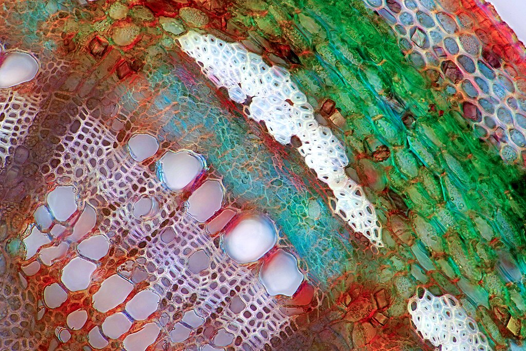
Transversal section through a rose stalk.
Summary
[edit]
| Description |
English: A big single vascular bundle is visible with xylem and phloem. White thick wall cells are sclerenchyma. External part of the stalk up to epidermis is visible too. This micrograph was taken in polarized light. This is fresh unstained material cut in fingers with shaving blade. English: Transversal section through a rose stalk |
| Date | |
| Source | Own work |
| Author | Misiek1962 |
Licensing
[edit]
I, the copyright holder of this work, hereby publish it under the following license:
This file is licensed under the Creative Commons Attribution 4.0 International license.
- You are free:
- to share – to copy, distribute and transmit the work
- to remix – to adapt the work
- Under the following conditions:
- attribution – You must give appropriate credit, provide a link to the license, and indicate if changes were made. You may do so in any reasonable manner, but not in any way that suggests the licensor endorses you or your use.
| This file was uploaded as part of Wiki Science Competition 2021. |
File history
Click on a date/time to view the file as it appeared at that time.
| Date/Time | Thumbnail | Dimensions | User | Comment | |
|---|---|---|---|---|---|
| current | 12:21, 1 December 2021 | | 6,667 × 4,453 (33.72 MB) | Misiek1962 (talk | contribs) | Uploaded own work with UploadWizard |
You cannot overwrite this file.
File usage on Commons
There are no pages that use this file.