File:Representative stem-group chordates and comparisons with Pikaia.png
Original file (3,321 × 1,810 pixels, file size: 9.58 MB, MIME type: image/png)
Captions
Captions
Summary
[edit]
| Description |
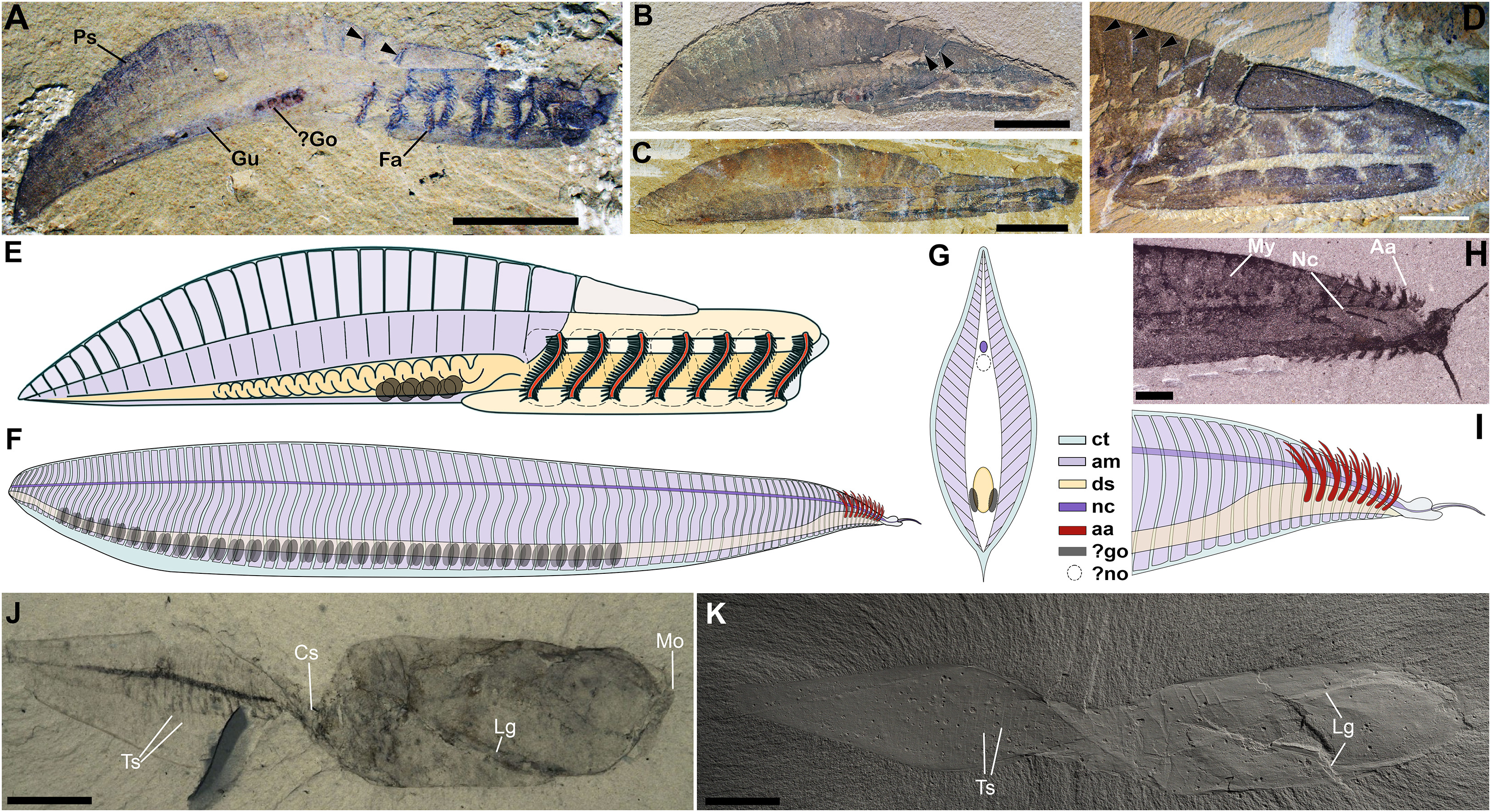
English: Figure 3 Representative stem-group chordates and comparisons with Pikaia
Hide caption (A–E) Yunnanozoon. (A) RCCBYU 10310, showing gut tube, posterior segments, and filamentous arches. (B) YKLP 13005. (C) YKLP 13032. (D) YKLP 13016. Black arrows denote separation between dorsal “segments.” (E) Schematic anatomical reconstruction after Cong et al. and Tian et al. (F) Pikaia, schematic reconstruction illustrating dorsoventrally inverted anatomical reinterpretation. (G) Hypothetical cross-sections of trunk segments in Pikaia3 under the new dorsoventrally inverted interpretation, showing dorsal and ventral “fins.”3 Color legend on the right applies to (E)–(I). (H) USNM 198688, detail of the anterior region showing bilobed head, anterior appendages, pharyngeal cavity, and dorsal nerve cord. (I) Schematic reconstruction of the anterior region of Pikaia under the new anatomical interpretation, showing the anterior appendages as dorsally directed gills. (J and K) Banffia constricta. (J) ROMIP 49914. (K) ROMIP 49898, photographed under ammonium chloride sublimate to highlight relief of lateral grooves and segmental boundaries. RCCBYU, Research Center of Chengjiang Biota, Yunnan University, Kunming; YKLP, Yunnan Key Laboratory for Palaeobiology, Kunming; USNM, Department of Paleobiology, National Museum of Natural History, Smithsonian Institution; ROMIP, Royal Ontario Museum, Invertebrate Palaeontology; Ps, posterior segments; Gu, gut; ?Go, possible gonads; Fa, filamentous arches; ct, connective tissue, integument; am, axial musculature; ds, digestive system; nc, dorsal nerve cord; aa, anterior appendages; ?no, hypothetical notochord; My, myomeres; Ph, pharynx; Lg, lateral grooves; Cs, constriction; Ts, tail segments; Mo, mouth opening. Scale bars, 5 mm (A, C, and D); 10 mm (B, J, and K); 1 mm (H). |
| Date | |
| Source | A new interpretation of Pikaia reveals the origins of the chordate body plan https://www.cell.com/current-biology/fulltext/S0960-9822(24)00669-9 |
| Author | Giovanni Mussini, M. Paul Smith, Jakob Vinther, Imran A. Rahman, Duncan J.E. Murdock, David A.T. Harper, Frances S. Dunn |
Licensing
[edit]
- You are free:
- to share – to copy, distribute and transmit the work
- to remix – to adapt the work
- Under the following conditions:
- attribution – You must give appropriate credit, provide a link to the license, and indicate if changes were made. You may do so in any reasonable manner, but not in any way that suggests the licensor endorses you or your use.
File history
Click on a date/time to view the file as it appeared at that time.
| Date/Time | Thumbnail | Dimensions | User | Comment | |
|---|---|---|---|---|---|
| current | 16:25, 12 June 2024 | | 3,321 × 1,810 (9.58 MB) | Ta-tea-two-te-to (talk | contribs) | Uploaded a work by Giovanni Mussini, M. Paul Smith, Jakob Vinther, Imran A. Rahman, Duncan J.E. Murdock, David A.T. Harper, Frances S. Dunn from A new interpretation of Pikaia reveals the origins of the chordate body plan https://www.cell.com/current-biology/fulltext/S0960-9822(24)00669-9 with UploadWizard |
You cannot overwrite this file.
File usage on Commons
The following 5 pages use this file: