File:Propagation of action potential along myelinated nerve fiber.png
Original file (3,554 × 1,973 pixels, file size: 777 KB, MIME type: image/png)
Captions
Captions
Summary
[edit]| Description |
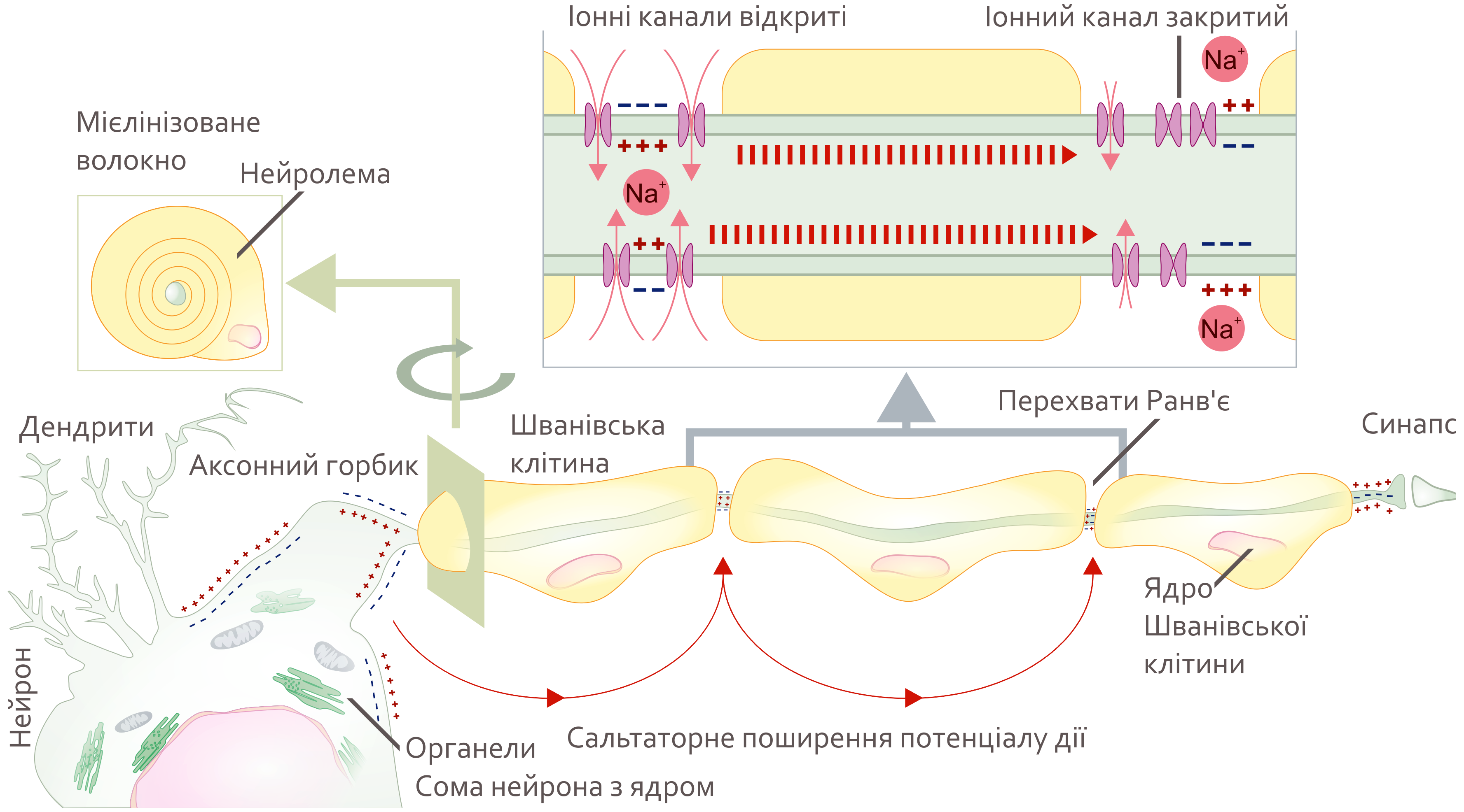
Українська: Цей файл в конкурсі участі не бере і не буде оцінюватися (бо автор – організатор і член журі конкурсу). Проте зображення потрібне для ілюстрації деяких статей, і автор давно хотіла його зробити – а тут такий гарний привід =) Опис зображення: Схематичне зображення поширення потенціалу дії через мієлінізоване волокно периферичної нервової системи. English: This image is made by one of the local juries of the contest. The image is not part of the contest, and will not participate in the competition and will not be rated. But it was helluva fun to make =) and some articles really need illustrations. Image description: Schematic representation of the action potential propagation through myelinated nerve fiber of peripheral nervous system. From axon hillock of neuron body (soma) action potential propagates from one unmyelinated fiber part to next one. The unmyelinated parts of the nerve fiber are nodes of Ranvier. This way of action potential propagation is called saltatory conduction (red arrows in the diagram) Ion channels open, allow potassium ions to enter the cell leading to membrane depolarization and generation of action potential. Myelination of nerve fibers in the peripheral nervous system is achieved by Schwann cells wrapping around an axon part (cross section). The nucleus and most of the Schwan cell cytoplasm are contained in the outer most layer called neurilemma. Own work loosely based on Raphael Alya R., Talbot William S. (2011). "New Insights into Signaling During Myelination in Zebrafish". Current topics in developmental biology 97: 1–19. DOI:10.1016/B978-0-12-385975-4.00007-3. ISSN 00702153.Min Y., Kristiansen K., Boggs J. M. at al (2009). "Interaction forces and adhesion of supported myelin lipid bilayers modulated by myelin basic protein". Proceedings of the National Academy of Sciences 106 (9): 3154–3159. DOI:10.1073/pnas.0813110106. ISSN 0027-8424. |
| Date | |
| Source | Own work |
| Author | Helixitta |
| Other versions |
|

Licensing
[edit]
- You are free:
- to share – to copy, distribute and transmit the work
- to remix – to adapt the work
- Under the following conditions:
- attribution – You must give appropriate credit, provide a link to the license, and indicate if changes were made. You may do so in any reasonable manner, but not in any way that suggests the licensor endorses you or your use.
- share alike – If you remix, transform, or build upon the material, you must distribute your contributions under the same or compatible license as the original.
| This image was uploaded as part of European Science Photo Competition 2015. |
File history
Click on a date/time to view the file as it appeared at that time.
| Date/Time | Thumbnail | Dimensions | User | Comment | |
|---|---|---|---|---|---|
| current | 18:32, 6 October 2015 | | 3,554 × 1,973 (777 KB) | Helixitta (talk | contribs) | minor changes. |
| 18:41, 2 October 2015 |  | 3,531 × 1,956 (790 KB) | Helixitta (talk | contribs) | User created page with UploadWizard |
You cannot overwrite this file.
File usage on Commons
The following 3 pages use this file:
File usage on other wikis
The following other wikis use this file: