File:Potamolithus santiagensis (10.5852-ejt.2019.524) Figure 3.png
Jump to navigation
Jump to search

Size of this preview: 547 × 599 pixels. Other resolutions: 219 × 240 pixels | 438 × 480 pixels | 701 × 768 pixels | 935 × 1,024 pixels | 1,891 × 2,072 pixels.
Original file (1,891 × 2,072 pixels, file size: 2.59 MB, MIME type: image/png)
Captions
Captions
Add a one-line explanation of what this file represents
Summary
[edit]_Figure_3.png&action=edit§ion=1)
| Description |
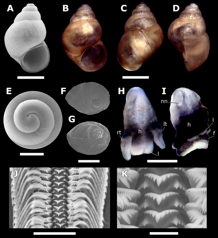
English: Fig. 3. Potamolithus santiagensis (Biese, 1944) comb. nov., Yeso Spring, Chile. A. Shell imaged using SEM. B–D. Shell of the same specimen photographed under a stereo microscope (frontal, dorsal, lateral views). E. Protoconch. F–G. Opercula of two specimens (outer, inner sides, respectively). H. Head of a female. I. Head of another female having a nuchal node. J. Anterior-central section of radular ribbon. K. Central teeth. Abbreviations: f = foot; h = head; l = lip; lt = left tentacle; nn = nuchal node; rt = right tentacle. Scale bars: A–D = 1.0 mm; E = 250 μm; F–G = 500 μm; H–I = 0.5 mm; J = 50 μm; K = 10 μm. |
| Date | |
| Source | https://dx.doi.org/10.5852%2Fejt.2019.524 |
| Author | Collado, G.A., Aguayo, K.P., Cazzaniga, N.J., Gutiérrez Gregoric, D.E., de Lucía, M., Haase, M. 2019. Systematic evaluation of cryptic freshwater snails from central Chile, including the enigmatic Littoridina santiagensis (Gastropoda, Truncatelloidea). European Journal of Taxonomy. 90(2): 133-146. DOI: 10.5852/ejt.2019.524 |
| Permission (Reusing this file) |
This file is licensed under the Creative Commons Attribution 4.0 International license.
|
File history
Click on a date/time to view the file as it appeared at that time.
| Date/Time | Thumbnail | Dimensions | User | Comment | |
|---|---|---|---|---|---|
| current | 12:29, 23 March 2021 | | 1,891 × 2,072 (2.59 MB) | Christian Ferrer (talk | contribs) | {{Information | description = {{en|1=Fig. 3. ''Potamolithus santiagensis'' (Biese, 1944) comb. nov., Yeso Spring, Chile. A. Shell imaged using SEM. B–D. Shell of the same specimen photographed under a stereo microscope (frontal, dorsal, lateral views). E. Protoconch. F–G. Opercula of two specimens (outer, inner sides, respectively). H. Head of a female. I. Head of another female having a nuchal node. J. Anterior-central section of radular ribbon. K. Central teeth. Abbreviations: f = foot; h... |
You cannot overwrite this file.
File usage on Commons
The following page uses this file:
File usage on other wikis
The following other wikis use this file:
- Usage on www.wikidata.org
_Figure_3.png&oldid=545532024)