File:Pipe bomb 05.jpg
Jump to navigation
Jump to search
No higher resolution available.
Pipe_bomb_05.jpg (744 × 261 pixels, file size: 101 KB, MIME type: image/jpeg)
Captions
Captions
Add a one-line explanation of what this file represents
| Description |
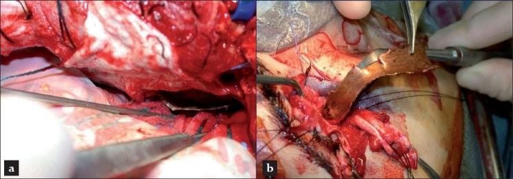
English: Intraoperative situs (a) intracranial view from above into the middle cranial fossa with cottonoids retracting the resection margin of the temporal lobe and (b) view onto the right supraorbital ridge and orbit during evacuation of the foreign body. (a)
Deutsch: Foto während der Entfernung eines metallischen Fremdkörpers einer Rohrbombe aus dem Gehirn eines Patienten. Intrakraniellen Blick von oben in die mittlere Schädelgrube mit Hirnwatten wird der Fremdkörper über den Resektionsrand des Temporallappens gezogen. (b) Blick auf den Hinterhauptfortsatz während der Entfernung des Fremdkörpers. |
| Date | Published: 25 December 2010 |
| Source | Kasper EM, Luedi MM, Zinn PO, Rubin PA, Chen C. Retained transorbital foreign body with intracranial extension after pipe bomb explosion. Surg Neurol Int 2010;1:94 doi:10.4103/2152-7806.74241 PMID 21246061 |
| Author | Ekkehard M Kasper, Markus M Luedi, Pascal O Zinn, Peter A.D Rubin, Clark Chen |
I, the copyright holder of this work, hereby publish it under the following license:
This file is licensed under the Creative Commons Attribution 2.0 Generic license.
- You are free:
- to share – to copy, distribute and transmit the work
- to remix – to adapt the work
- Under the following conditions:
- attribution – You must give appropriate credit, provide a link to the license, and indicate if changes were made. You may do so in any reasonable manner, but not in any way that suggests the licensor endorses you or your use.
File history
Click on a date/time to view the file as it appeared at that time.
| Date/Time | Thumbnail | Dimensions | User | Comment | |
|---|---|---|---|---|---|
| current | 20:22, 3 January 2013 | 744 × 261 (101 KB) | Kuebi (talk | contribs) | {{Information |Description={{en|Intraoperative situs (a) intracranial view from above into the middle cranial fossa with cottonoids retracting the resection margin of the temporal lobe and (b) view onto the right supraorbital ridge and orbit during eva... |
You cannot overwrite this file.
File usage on Commons
There are no pages that use this file.